


anti-alpha Synuclein antibody [2A7]
![anti-alpha Synuclein antibody [2A7]](/upload/image/products/ARG43207_IHC-Fr_3_210_205.jpg)
![anti-alpha Synuclein antibody [2A7]](/upload/image/products/ARG43207_IHC-Fr_1.jpg)
![anti-alpha Synuclein antibody [2A7]](/upload/image/products/ARG43207_WB_1.jpg)
![anti-alpha Synuclein antibody [2A7]](/upload/image/products/ARG43207_IHC-P_1.jpg)
![anti-alpha Synuclein antibody [2A7]](/upload/image/products/ARG43207_IHC-Fr_2.jpg)
![anti-alpha Synuclein antibody [2A7]](/upload/image/products/ARG43207_IHC-Fr_3.jpg)
Key features and details
- 产品描述:
- 反应物种:
- 应用:
- 宿主:
- 克隆:
- 克隆号:
- 同位型:
- 靶点名称:
- 抗原物种:
-
Brand:

 Product Details
| 产品描述 | Mouse Monoclonal antibody [2A7] recognizes alpha Synuclein |
|---|---|
| 反应物种 | Hu, Ms, Rat, Cow, Pig |
| 应用 | ICC/IF, IHC-Fr, IHC-P, WB |
| 宿主 | Mouse |
| 克隆 | Monoclonal |
| 克隆号 | 2A7 |
| 同位型 | IgG1 |
| 靶点名称 | alpha Synuclein |
| 抗原物种 | Human |
| 抗原 | Recombinant full length Human alpha synuclein protein. |
| 偶联标记 | Un-conjugated |
| 別名 | Non-A4 component of amyloid precursor; Alpha-synuclein; PARK4; PARK1; PD1; NACP; Non-A beta component of AD amyloid |
| 应用建议 |
| ||||||||||
|---|---|---|---|---|---|---|---|---|---|---|---|
| 应用说明 | * The dilutions indicate recommended starting dilutions and the optimal dilutions or concentrations should be determined by the scientist. | ||||||||||
| 阳性对照 | Rat brain and Rat spinal cord | ||||||||||
| 实际分子量 | ~ 17 kDa |
| 形式 | Liquid |
|---|---|
| 纯化 | Purified |
| 缓冲液 | PBS, 5 mM Sodium azide and 50% Glycerol. |
| 抗菌剂 | 5 mM Sodium azide |
| 稳定剂 | 50% Glycerol |
| 浓度 | 1 mg/ml |
| 存放说明 | For continuous use, store undiluted antibody at 2-8°C for up to a week. For long-term storage, aliquot and store at -20°C. Storage in frost free freezers is not recommended. Avoid repeated freeze/thaw cycles. Suggest spin the vial prior to opening. The antibody solution should be gently mixed before use. |
| 注意事项 | For laboratory research only, not for drug, diagnostic or other use. |
| 数据库连接 | |
|---|---|
| 基因名称 | SNCA |
| 全名 | synuclein, alpha (non A4 component of amyloid precursor) |
| 背景介绍 | Alpha-synuclein is a member of the synuclein family, which also includes beta- and gamma-synuclein. Synucleins are abundantly expressed in the brain and alpha- and beta-synuclein inhibit phospholipase D2 selectively. SNCA may serve to integrate presynaptic signaling and membrane trafficking. Defects in SNCA have been implicated in the pathogenesis of Parkinson disease. SNCA peptides are a major component of amyloid plaques in the brains of patients with Alzheimer's disease. Alternatively spliced transcripts encoding different isoforms have been identified for this gene. [provided by RefSeq, Feb 2016] |
| 生物功能 | Neuronal protein that plays several roles in synaptic activity such as regulation of synaptic vesicle trafficking and subsequent neurotransmitter release. Participates as a monomer in synaptic vesicle exocytosis by enhancing vesicle priming, fusion and dilation of exocytotic fusion pores (PubMed:28288128, PubMed:30404828). Mechanistically, acts by increasing local Ca(2+) release from microdomains which is essential for the enhancement of ATP-induced exocytosis (PubMed:30404828). Acts also as a molecular chaperone in its multimeric membrane-bound state, assisting in the folding of synaptic fusion components called SNAREs (Soluble NSF Attachment Protein REceptors) at presynaptic plasma membrane in conjunction with cysteine string protein-alpha/DNAJC5 (PubMed:20798282). This chaperone activity is important to sustain normal SNARE-complex assembly during aging (PubMed:20798282). Plays also a role in the regulation of the dopamine neurotransmission by associating with the dopamine transporter (DAT1) and thereby modulating its activity (PubMed:26442590). [UniProt] |
| 细胞定位 | Cytoplasm, cytosol. Membrane. Nucleus. Cell junction, synapse. Secreted. Note=Membrane-bound in dopaminergic neurons. [UniProt] |
| 预测分子量 | 14 kDa |
| 翻译后修饰 | Phosphorylated, predominantly on serine residues. Phosphorylation by CK1 appears to occur on residues distinct from the residue phosphorylated by other kinases. Phosphorylation of Ser-129 is selective and extensive in synucleinopathy lesions. In vitro, phosphorylation at Ser-129 promoted insoluble fibril formation. Phosphorylated on Tyr-125 by a PTK2B-dependent pathway upon osmotic stress. Hallmark lesions of neurodegenerative synucleinopathies contain alpha-synuclein that is modified by nitration of tyrosine residues and possibly by dityrosine cross-linking to generated stable oligomers. Ubiquitinated. The predominant conjugate is the diubiquitinated form (By similarity). Acetylation at Met-1 seems to be important for proper folding and native oligomeric structure. [UniProt] |
ARG43207 anti-alpha Synuclein antibody [2A7] IHC-Fr image
Immunohistochemistry: Frozen section of Rat cerebellum tissue stained with ARG43207 anti-alpha Synuclein antibody [2A7] (red) at 1:1000 dilution, and costained with anti-GFAP antibody (green) at 1:5000 dilution. Hoechst (blue) for nuclear staining. Sample preparation: Following transcardial perfusion of Rat with 4% paraformaldehyde, brain was post fixed for 24 hours, cut to 45 µM, and free-floating sections were stained with above antibodies.
The alpha synuclein protein is concentrated in synaptic regions, while the GFAP antibody stains the filamentous cytoskeleton of Bergmann glia and astrocytic cells.
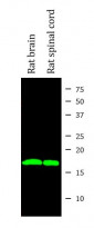
ARG43207 anti-alpha Synuclein antibody [2A7] WB image
Western blot: Rat brain and Rat spinal cord lysates stained with ARG43207 anti-alpha Synuclein antibody [2A7] (green) at 1:1000 dilution.
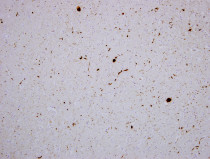
ARG43207 anti-alpha Synuclein antibody [2A7] IHC-P image
Immunohistochemistry: Paraffin-embedded cortex tissue of a patient with Parkinson's disease. Tissue section was stained with ARG43207 anti-alpha Synuclein antibody [2A7] at 1:1000 dilution. Antibody revealed with HRP and DAB.
The Lewy bodies and other typical inclusions of Parkinson's disease are seen in brown.
ARG43207 anti-alpha Synuclein antibody [2A7] IHC-Fr image
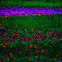
Immunohistochemistry: Frozen section of Rat hippocampus tissue stained with ARG43207 anti-alpha Synuclein antibody [2A7] (green) at 1:1000 dilution, and costained with anti-MeCP2 antibody (red) at 1:2000 dilution. DAPI (blue) for nuclear staining. Sample preparation: Following transcardial perfusion of Rat with 4% paraformaldehyde, brain was post fixed for 1 hour, cut to 45 µM, and free-floating sections were stained with above antibodies.
The alpha synuclein protein is concentrated in synaptic regions, and the MeCP2 antibody stains the nuclei of neuronal cells.
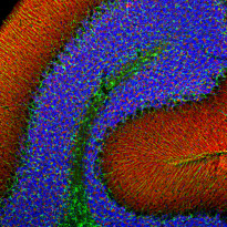
ARG43207 anti-alpha Synuclein antibody [2A7] IHC-Fr image
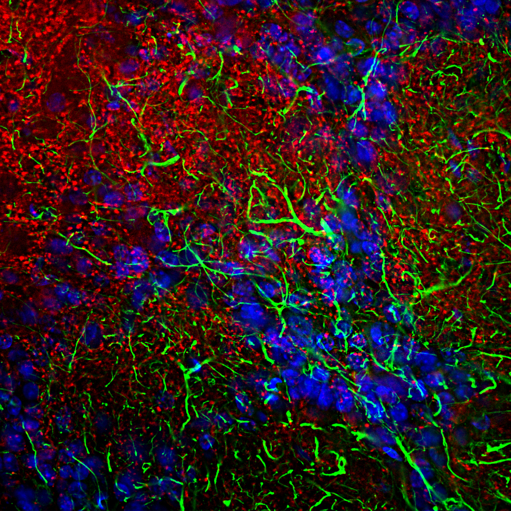
Immunohistochemistry: Frozen section of Rat olfactory bulb tissue stained with ARG43207 anti-alpha Synuclein antibody [2A7] (red) at 1:1000 dilution, and costained with anti-GFAP antibody (green) at 1:5000 dilution. DAPI (blue) for nuclear staining. Sample preparation: Following transcardial perfusion of Rat with 4% paraformaldehyde, brain was post fixed for 24 hours, cut to 45 µM, and free-floating sections were stained with above antibodies.
The alpha synuclein protein is concentrated in synaptic regions, while the GFAP antibody stains the filamentous backbone of astroglial cells.


 New Products


