


anti-beta III Tubulin antibody [TU-20]
![anti-beta III Tubulin antibody [TU-20]](/upload/image/products/ARG62683_SheetRevABC_160531.jpg)
![anti-beta III Tubulin antibody [TU-20]](/upload/image/products/ARG62683_IHC-P_1_201117.jpg)
![anti-beta III Tubulin antibody [TU-20]](/upload/image/products/TU-20_1.jpg)
![anti-beta III Tubulin antibody [TU-20]](/upload/image/products/圖片2.jpg)
![anti-beta III Tubulin antibody [TU-20]](/upload/image/products/TU-20_2.jpg)
![anti-beta III Tubulin antibody [TU-20]](/upload/image/products/TU-20_3.jpg)
Key features and details
- 产品描述:
- 反应物种:
- 应用:
- 特异性:
- 宿主:
- 克隆:
- 克隆号:
- 同位型:
- 靶点名称:
-
Brand:

 Product Details
| 产品描述 | Mouse Monoclonal antibody [TU-20] recognizes beta III Tubulin |
|---|---|
| 反应物种 | Hu, Ms, Rat, Dog, Pig |
| 应用 | FACS, ICC/IF, IHC-Fr, IHC-P, WB |
| 特异性 | The clone TU-20 recognizes C-terminal peptide sequence ESESQGPK (aa 441-448) of neuron-specific human beta III Tubulin. |
| 宿主 | Mouse |
| 克隆 | Monoclonal |
| 克隆号 | TU-20 |
| 同位型 | IgG1 |
| 靶点名称 | beta III Tubulin |
| 抗原 | Peptide (C) 441-448 coupled to maleimide-activated keyhole limpet hemocyanin via cysteine added to the N-terminus of the neuron-specific peptide. |
| 偶联标记 | Un-conjugated |
| 別名 | CDCBM1; Tubulin beta-4 chain; Tubulin beta-3 chain; CFEOM3A; Tubulin beta-III; TUBB4; CDCBM; CFEOM3; FEOM3; beta-4 |
| 应用建议 |
| ||||||||||||
|---|---|---|---|---|---|---|---|---|---|---|---|---|---|
| 应用说明 | WB: In reducing conditions, incubated for 90 min. Positive control: Porcine brain lysate; Negative control: HPB-ALL human peripheral blood leukemia cell line. Sample preparation: Mix lysate with reducing Laemmli SDS-PAGE sample buffer. IHC-P: Staining technique: Standard ABC technique (DAB+). Antigen Retrieval: Heat the tissue section in 10 mM Citrate buffer (pH 6.0) or pretreat samples with 0.1% pepsin (trypsin) in 0.1M HCl for 30 min at RT. * The dilutions indicate recommended starting dilutions and the optimal dilutions or concentrations should be determined by the scientist. | ||||||||||||
| 阳性对照 | Positive tissue: neuronal tissue Immunocytochemistry: Positive material: Neuro2a mouse neuroblastoma cell line |
| 形式 | Liquid |
|---|---|
| 纯化 | Purified by ammonium sulphate and caprylic acid precipitation |
| 纯度 | > 95% (by SDS-PAGE) |
| 缓冲液 | PBS (pH 7.4) and 15 mM Sodium azide |
| 抗菌剂 | 15 mM Sodium azide |
| 浓度 | 1 mg/ml |
| 存放说明 | For continuous use, store undiluted antibody at 2-8°C for up to a week. For long-term storage, aliquot and store at -20°C or below. Storage in frost free freezers is not recommended. Avoid repeated freeze/thaw cycles. Suggest spin the vial prior to opening. The antibody solution should be gently mixed before use. |
| 注意事项 | For laboratory research only, not for drug, diagnostic or other use. |
| 数据库连接 | |
|---|---|
| 基因名称 | TUBB3 |
| 全名 | tubulin, beta 3 class III |
| 背景介绍 | The beta III-Tubulin isoform is present dominantly in cells of neuronal origin and it is one of the earliest markers of neuronal differentiation. It is regarded as a specific probe for the cells of neuronal origin as well as for the tumours originating from these cells. The betaIII-Tubulin is most abundant in cells of neuronal origin, but was also detected in Sertoli cells of the testis and transiently in non-neuronal embryonic tissues. |
| 生物功能 | Tubulin is the major constituent of microtubules. It binds two moles of GTP, one at an exchangeable site on the beta chain and one at a non-exchangeable site on the alpha chain. TUBB3 plays a critical role in proper axon guidance and mantainance. [UniProt] |
| 产品亮点 | Related Antibody Duos and Panels: ARG30010 Neuronal Cytoskeletal Antibody Duo (NF-L, TUBBIII) ARG30301 Neurite Marker Antibody Duo Related products: beta III Tubulin antibodies; beta III Tubulin Duos / Panels; Anti-Mouse IgG secondary antibodies; Related news: Stem cell and the regenerative medicine: Ready for the patients Neuronal Development Marker Choose the Best ZIKA Virus Antibodies Fight microcephaly with arigo Astrocyte-to-neuron conversion for Parkinson's disease treatment |
| 研究领域 | Controls and Markers antibody; Neuroscience antibody; Signaling Transduction antibody; Neuron Development Study antibody; Neuronal Cytoskeletal antibody; Neurite Marker antibody |
| 预测分子量 | 50 kDa |
| 翻译后修饰 | Some glutamate residues at the C-terminus are polyglutamylated, resulting in polyglutamate chains on the gamma-carboxyl group (PubMed:26875866). Polyglutamylation plays a key role in microtubule severing by spastin (SPAST). SPAST preferentially recognizes and acts on microtubules decorated with short polyglutamate tails: severing activity by SPAST increases as the number of glutamates per tubulin rises from one to eight, but decreases beyond this glutamylation threshold (PubMed:26875866). Some glutamate residues at the C-terminus are monoglycylated but not polyglycylated due to the absence of functional TTLL10 in human. Monoglycylation is mainly limited to tubulin incorporated into axonemes (cilia and flagella). Both polyglutamylation and monoglycylation can coexist on the same protein on adjacent residues, and lowering glycylation levels increases polyglutamylation, and reciprocally. The precise function of monoglycylation is still unclear (Probable). Phosphorylated on Ser-172 by CDK1 during the cell cycle, from metaphase to telophase, but not in interphase. This phosphorylation inhibits tubulin incorporation into microtubules. |
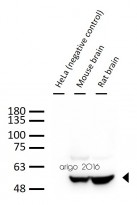
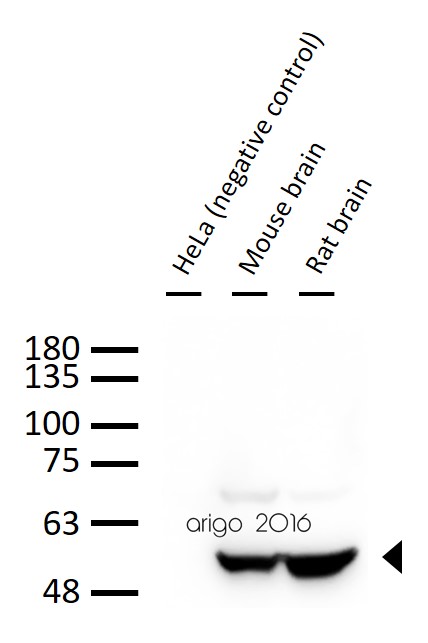
ARG62683 anti-beta III Tubulin antibody [TU-20] WB image
Western blot: 30 µg of 1) HeLa (negative control), 2) Mouse brain, and 3) Rat brain lysate stained with ARG62683 anti-beta III Tubulin antibody [TU-20] at 1:1000 dilution.
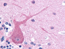
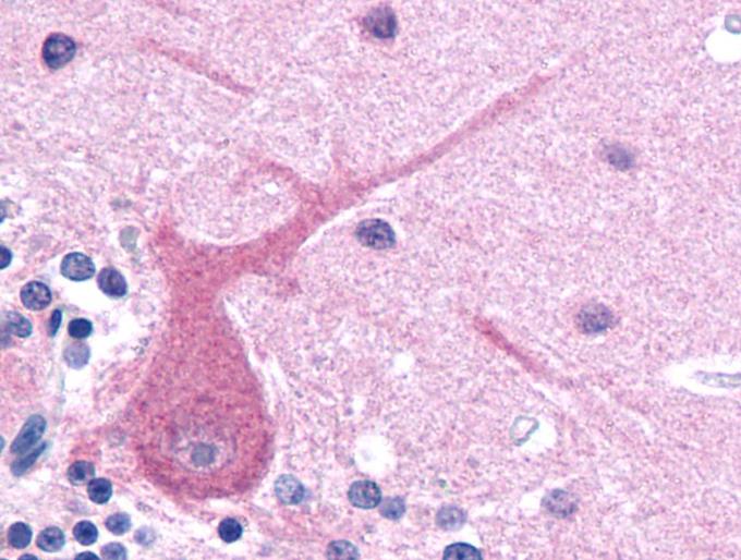
ARG62683 anti-beta III Tubulin antibody [TU-20] IHC-P image
Immunohistochemistry: Paraffin-embedded Human brain tissue stained with ARG62683 anti-beta III Tubulin antibody [TU-20].
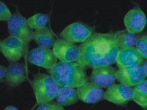
ARG62683 anti-beta III Tubulin antibody [TU-20] ICC/IF image
Immunofluorescence: Neuro2a mouse neuroblastoma cell stained with ARG62683 anti-beta III Tubulin antibody [TU-20] (green).
Cell nuclei was stained with DAPI (blue).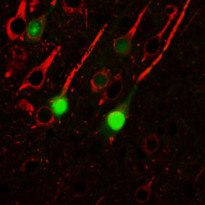
ARG62683 anti-beta III Tubulin antibody [TU-20] IHC-P image
Immunohistochemistry: Mouse brain stained with ARG62683 anti-beta III Tubulin antibody [TU-20].
ARG62683 anti-beta III Tubulin antibody [TU-20] ICC/IF image
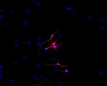
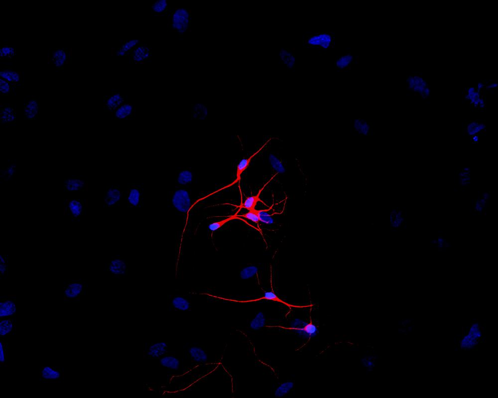
Immunofluorescence: P-19 mouse embryonal carcinoma cells stimulated to neuronal differentiation by retinoic acid stained with ARG62683 anti-beta III Tubulin antibody [TU-20] (red).
Cell nuclei was stained with DAPI (blue).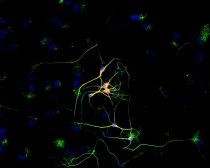
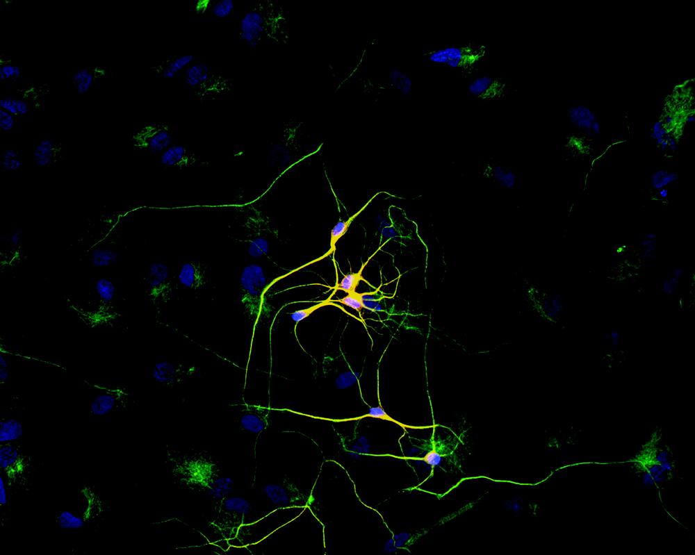
ARG62683 anti-beta III Tubulin antibody [TU-20] ICC/IF image
Immunofluorescence: P-19 mouse embryonal carcinoma cell line stimulated to neuronal differentiation by retinoic acid co-stained with stained with ARG62683 anti-beta III Tubulin antibody [TU-20] (red) and anti-beta-tubulin (green)
Superposition of red and green colours provided yellow staining. Nuclei were stained with DNA-binding dye (blue).


Magnetic stirring with iron oxide nanospinners accretes neurotoxic Aβ42 oligomers into phagocytic clearable plaques for Alzheimer's disease treatment
ICC/IF / Mouse


BAF45D Downregulation in Spinal Cord Ependymal Cells Following Spinal Cord Injury in Adult Rats and Its Potential Role in the Development of Neuronal Lesions.
WB, IHC-Fr / Rat
New Products


