


anti-EphA2 antibody

Key features and details
- 产品描述:
- 反应物种:
- 应用:
- 宿主:
- 克隆:
- 同位型:
- 靶点名称:
- 抗原物种:
- 抗原:
-
Brand:

 Product Details
| 产品描述 | Rabbit Polyclonal antibody recognizes EphA2 |
|---|---|
| 反应物种 | Hu, Ms |
| 应用 | FACS, ICC/IF, WB |
| 宿主 | Rabbit |
| 克隆 | Polyclonal |
| 同位型 | IgG |
| 靶点名称 | EphA2 |
| 抗原物种 | Human |
| 抗原 | Recombinant protein corresponding to M851-N970 of Human EphA2. |
| 偶联标记 | Un-conjugated |
| 別名 | CTRCT6; Ephrin type-A receptor 2; Tyrosine-protein kinase receptor ECK; Epithelial cell kinase; ECK; ARCC2; CTPA; CTPP1; EC 2.7.10.1 |
| 应用建议 |
| ||||||||
|---|---|---|---|---|---|---|---|---|---|
| 应用说明 | * The dilutions indicate recommended starting dilutions and the optimal dilutions or concentrations should be determined by the scientist. |
| 形式 | Liquid |
|---|---|
| 纯化 | Purified. |
| 缓冲液 | 0.9% NaCl, 0.2% Na2HPO4, 0.05% Sodium azide and 4% Trehalose. |
| 抗菌剂 | 0.05% Sodium azide |
| 稳定剂 | 4% Trehalose |
| 浓度 | 0.5 mg/ml |
| 存放说明 | For continuous use, store undiluted antibody at 2-8°C for up to a week. For long-term storage, aliquot and store at -20°C or below. Storage in frost free freezers is not recommended. Avoid repeated freeze/thaw cycles. Suggest spin the vial prior to opening. The antibody solution should be gently mixed before use. |
| 注意事项 | For laboratory research only, not for drug, diagnostic or other use. |
| 数据库连接 | |
|---|---|
| 基因名称 | EPHA2 |
| 全名 | EPH receptor A2 |
| 背景介绍 | This gene belongs to the ephrin receptor subfamily of the protein-tyrosine kinase family. EPH and EPH-related receptors have been implicated in mediating developmental events, particularly in the nervous system. Receptors in the EPH subfamily typically have a single kinase domain and an extracellular region containing a Cys-rich domain and 2 fibronectin type III repeats. The ephrin receptors are divided into 2 groups based on the similarity of their extracellular domain sequences and their affinities for binding ephrin-A and ephrin-B ligands. This gene encodes a protein that binds ephrin-A ligands. Mutations in this gene are the cause of certain genetically-related cataract disorders.[provided by RefSeq, May 2010] |
| 生物功能 | Receptor tyrosine kinase which binds promiscuously membrane-bound ephrin-A family ligands residing on adjacent cells, leading to contact-dependent bidirectional signaling into neighboring cells. The signaling pathway downstream of the receptor is referred to as forward signaling while the signaling pathway downstream of the ephrin ligand is referred to as reverse signaling. Activated by the ligand ephrin-A1/EFNA1 regulates migration, integrin-mediated adhesion, proliferation and differentiation of cells. Regulates cell adhesion and differentiation through DSG1/desmoglein-1 and inhibition of the ERK1/ERK2 (MAPK3/MAPK1, respectively) signaling pathway. May also participate in UV radiation-induced apoptosis and have a ligand-independent stimulatory effect on chemotactic cell migration. During development, may function in distinctive aspects of pattern formation and subsequently in development of several fetal tissues. Involved for instance in angiogenesis, in early hindbrain development and epithelial proliferation and branching morphogenesis during mammary gland development. Engaged by the ligand ephrin-A5/EFNA5 may regulate lens fiber cells shape and interactions and be important for lens transparency development and maintenance. With ephrin-A2/EFNA2 may play a role in bone remodeling through regulation of osteoclastogenesis and osteoblastogenesis. [UniProt] |
| 细胞定位 | Cell membrane. [UniProt] |
| 预测分子量 | 108 kDa |
| 翻译后修饰 | Autophosphorylates. Phosphorylated on tyrosine upon binding and activation by EFNA1. Phosphorylated residues Tyr-588 and Tyr-594 are required for binding VAV2 and VAV3 while phosphorylated residues Tyr-735 and Tyr-930 are required for binding PI3-kinase p85 subunit (PIK3R1, PIK3R2 or PIK3R3). These phosphorylated residues are critical for recruitment of VAV2 and VAV3 and PI3-kinase p85 subunit which transduce downstream signaling to activate RAC1 GTPase and cell migration. Dephosphorylation of Tyr-930 by PTPRF prevents the interaction of EPHA2 with NCK1. Phosphorylated at Ser-897 by PKB; serum-induced phosphorylation which targets EPHA2 to the cell leading edge and stimulates cell migration. Phosphorylation by PKB is inhibited by EFNA1-activated EPHA2 which regulates PKB activity via a reciprocal regulatory loop. Phosphorylated at Ser-897 in response to TNF by RPS6KA1 and RPS6KA3; RPS6KA-EPHA2 signaling pathway controls cell migration (PubMed:26158630). Phosphorylated at Ser-897 by PKA; blocks cell retraction induced by EPHA2 kinase activity (PubMed:27385333). Dephosphorylated by ACP1. Ubiquitinated by CHIP/STUB1. Ubiquitination is regulated by the HSP90 chaperone and regulates the receptor stability and activity through proteasomal degradation. ANKS1A prevents ubiquitination and degradation (By similarity). [UniProt] |
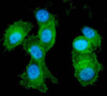
ARG58571 anti-EphA2 antibody ICC/IF image
Immunofluorescence: PC-3 cells were blocked with 10% goat serum and then stained with ARG58571 anti-EphA2 antibody (green) at 5 µg/ml dilution, overnight at 4°C. DAPI (blue) for nuclear staining.
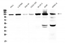
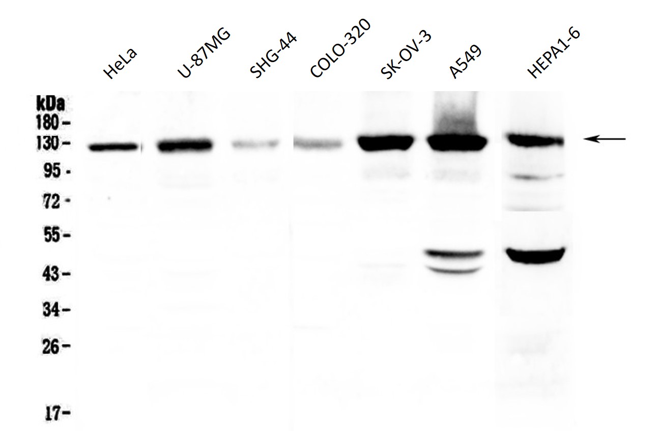
ARG58571 anti-EphA2 antibody WB image
Western blot: 50 µg of HeLa, U-87MG, SHG-44, COLO-320, SK-OV-3, A549 and HEPA1-6 cell lysates stained with ARG58571 anti-EphA2 antibody at 0.5 µg/ml, overnight at 4°C, under reducing conditions.

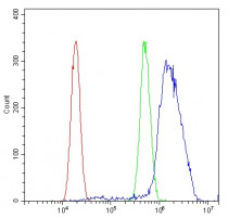
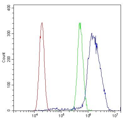
ARG58571 anti-EphA2 antibody FACS image
Flow Cytometry: A549 cells were blocked with 10% normal goat serum and then stained with ARG58571 anti-EphA2 antibody (blue) at 1 µg/10^6 cells for 30 min at 20°C, followed by incubation with DyLight®488 labelled secondary antibody. Isotype control antibody (green) was rabbit IgG (1 µg/10^6 cells) used under the same conditions. Unlabelled sample (red) was also used as a control.
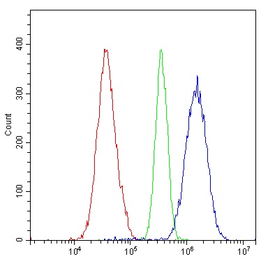
ARG58571 anti-EphA2 antibody FACS image
Flow Cytometry: U2OS cells were blocked with 10% normal goat serum and then stained with ARG58571 anti-EphA2 antibody (blue) at 1 µg/10^6 cells for 30 min at 20°C, followed by incubation with DyLight®488 labelled secondary antibody. Isotype control antibody (green) was rabbit IgG (1 µg/10^6 cells) used under the same conditions. Unlabelled sample (red) was also used as a control.


 New Products


