


anti-LCP1 / Plastin L antibody


Key features and details
- 产品描述:
- 反应物种:
- 应用:
- 宿主:
- 克隆:
- 同位型:
- 靶点名称:
- 抗原物种:
- 抗原:
-
Brand:

 Product Details
| 产品描述 | Rabbit Polyclonal antibody recognizes LCP1 / Plastin L |
|---|---|
| 反应物种 | Hu, Ms, Rat |
| 应用 | FACS, ICC/IF, IHC-P, WB |
| 宿主 | Rabbit |
| 克隆 | Polyclonal |
| 同位型 | IgG |
| 靶点名称 | LCP1 / Plastin L |
| 抗原物种 | Human |
| 抗原 | Recombinant protein corresponding to M1-K444 of Human LCP1. |
| 偶联标记 | Un-conjugated |
| 別名 | LC64P; L-PLASTIN; CP64; HEL-S-37; LCP-1; LPL; L-plastin; Lymphocyte cytosolic protein 1; Plastin-2; PLS2 |
| 应用建议 |
| ||||||||||
|---|---|---|---|---|---|---|---|---|---|---|---|
| 应用说明 | IHC-P: Antigen Retrieval: Heat mediation was performed in Citrate buffer (pH 6.0) for 20 min. * The dilutions indicate recommended starting dilutions and the optimal dilutions or concentrations should be determined by the scientist. | ||||||||||
| 实际分子量 | 70 kDa |
| 形式 | Liquid |
|---|---|
| 纯化 | Affinity purification with immunogen. |
| 缓冲液 | 0.2% Na2HPO4, 0.9% NaCl, 0.05% Sodium azide and 4% Trehalose. |
| 抗菌剂 | 0.05% Sodium azide |
| 稳定剂 | 4% Trehalose |
| 浓度 | 0.5 mg/ml |
| 存放说明 | For continuous use, store undiluted antibody at 2-8°C for up to a week. For long-term storage, aliquot and store at -20°C or below. Storage in frost free freezers is not recommended. Avoid repeated freeze/thaw cycles. Suggest spin the vial prior to opening. The antibody solution should be gently mixed before use. |
| 注意事项 | For laboratory research only, not for drug, diagnostic or other use. |
| 数据库连接 | |
|---|---|
| 基因名称 | LCP1 |
| 全名 | lymphocyte cytosolic protein 1 (L-plastin) |
| 背景介绍 | Plastins are a family of actin-binding proteins that are conserved throughout eukaryote evolution and expressed in most tissues of higher eukaryotes. In humans, two ubiquitous plastin isoforms (L and T) have been identified. Plastin 1 (otherwise known as Fimbrin) is a third distinct plastin isoform which is specifically expressed at high levels in the small intestine. The L isoform is expressed only in hemopoietic cell lineages, while the T isoform has been found in all other normal cells of solid tissues that have replicative potential (fibroblasts, endothelial cells, epithelial cells, melanocytes, etc.). However, L-plastin has been found in many types of malignant human cells of non-hemopoietic origin suggesting that its expression is induced accompanying tumorigenesis in solid tissues. [provided by RefSeq, Jul 2008] |
| 生物功能 | Actin-binding protein. Plays a role in the activation of T-cells in response to costimulation through TCR/CD3 and CD2 or CD28. Modulates the cell surface expression of IL2RA/CD25 and CD69. [UniProt] |
| 细胞定位 | Cytoplasm, cytoskeleton. Cell junction. Cell projection. Cell projection, ruffle membrane; Peripheral membrane protein; Cytoplasmic side. Note=Relocalizes to the immunological synapse between peripheral blood T-lymphocytes and antibody-presenting cells in response to costimulation through TCR/CD3 and CD2 or CD28 (PubMed:17294403). Associated with the actin cytoskeleton at membrane ruffles. Relocalizes to actin-rich cell projections upon serine phosphorylation (PubMed:16636079). [UniProt] |
| 预测分子量 | 70 kDa |
| 翻译后修饰 | Phosphorylated on a serine residue in response to costimulation through TCR/CD3 and CD2 or CD28. Serine phosphorylation promotes association with the actin cytoskeleton and targeting to peripheral cell projections. [UniProt] |

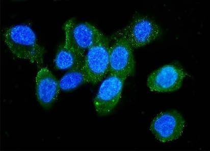
ARG40380 anti-LCP1 / Plastin L antibody ICC/IF image
Immunofluorescence: A431 cells were blocked with 10% goat serum and then stained with ARG40380 anti-LCP1 / Plastin L antibody (green) at 2 µg/ml dilution, overnight at 4°C. DAPI (blue) for nuclear staining.
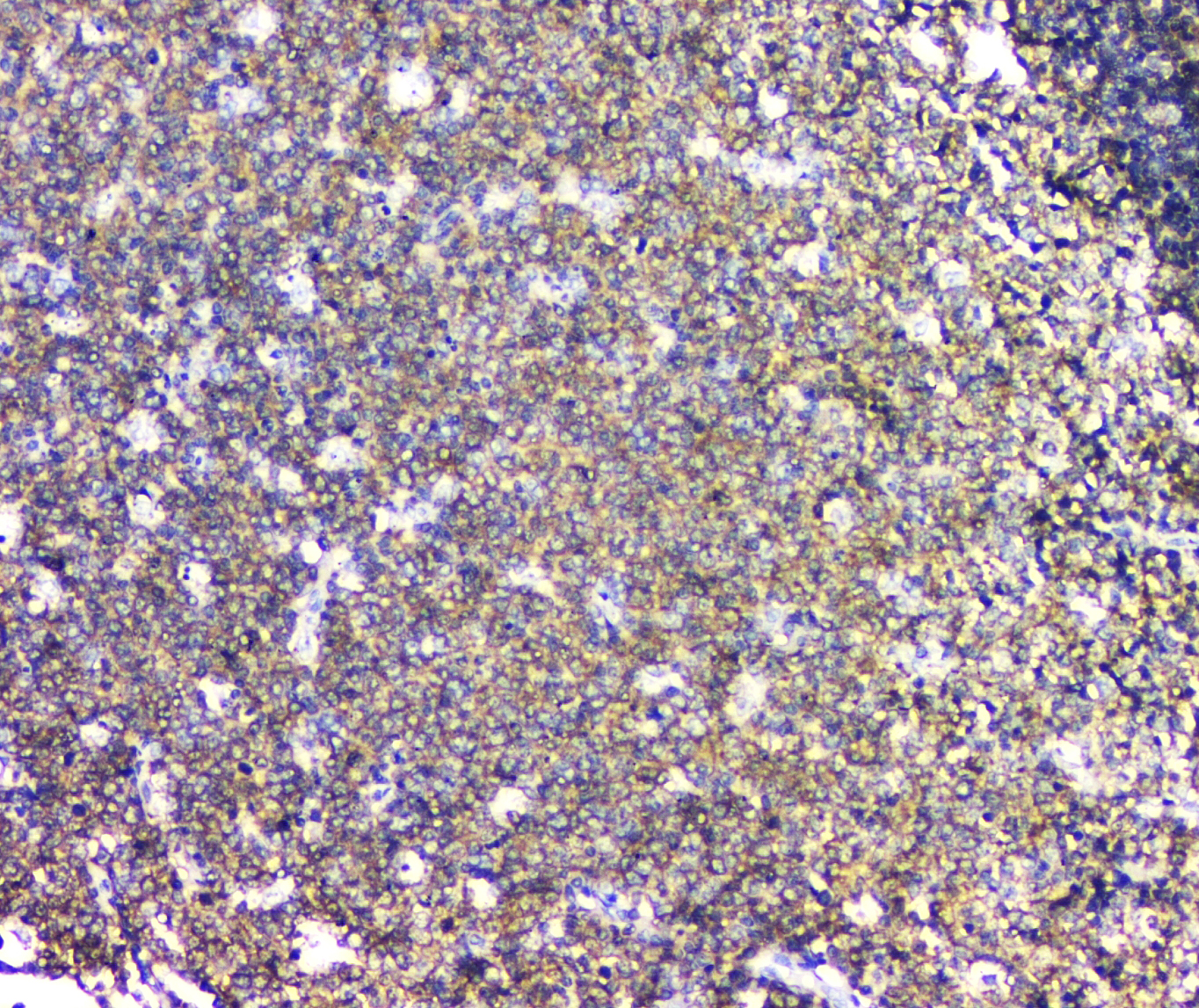
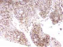
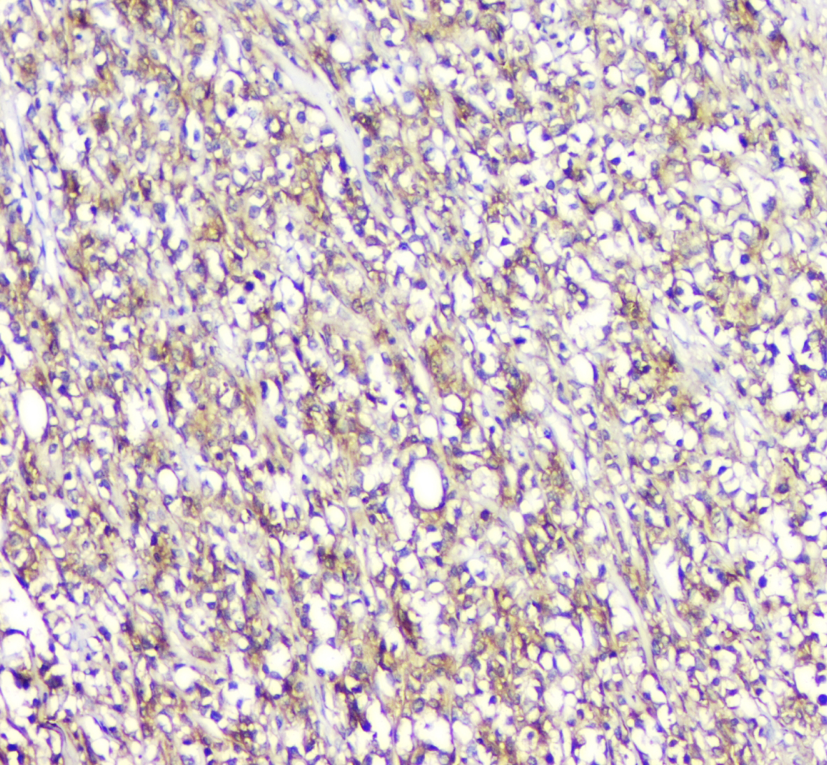
ARG40380 anti-LCP1 / Plastin L antibody IHC-P image
Immunohistochemistry: Paraffin-embedded Human B lymphocytic tumor tissue. Antigen Retrieval: Heat mediation was performed in Citrate buffer (pH 6.0, epitope retrieval solution) for 20 min. The tissue section was blocked with 10% goat serum. The tissue section was then stained with ARG40380 anti-LCP1 / Plastin L antibody at 1 µg/ml, overnight at 4°C.
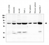
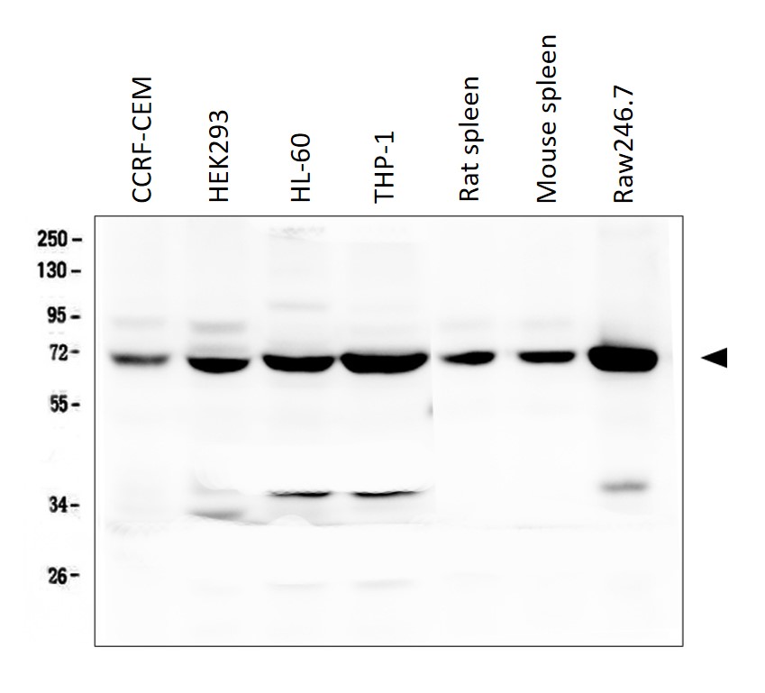
ARG40380 anti-LCP1 / Plastin L antibody WB image
Western blot: 50 µg of samples under reducing conditions. Human CCRF-CEM, HEK293, HL-60, THP-1, Rat spleen, Mouse spleen and Raw246.7 cell lysates stained with ARG40380 anti-LCP1 / Plastin L antibody at 0.5 µg/ml, overnight at 4°C.
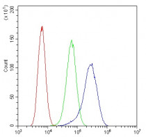
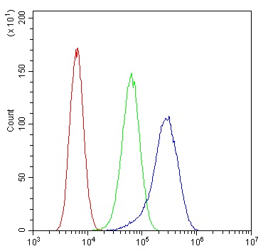
ARG40380 anti-LCP1 / Plastin L antibody FACS image
Flow Cytometry: U2OS cells were blocked with 10% normal goat serum and then stained with ARG40380 anti-LCP1 / Plastin L antibody (blue) at 1 µg/10^6 cells for 30 min at 20°C, followed by incubation with DyLight®488 labelled secondary antibody. Isotype control antibody (green) was rabbit IgG (1 µg/10^6 cells) used under the same conditions. Unlabelled sample (red) was also used as a control.
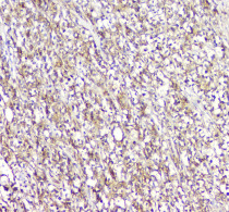
ARG40380 anti-LCP1 / Plastin L antibody IHC-P image
Immunohistochemistry: Paraffin-embedded Human tonsil tissue. Antigen Retrieval: Heat mediation was performed in Citrate buffer (pH 6.0, epitope retrieval solution) for 20 min. The tissue section was blocked with 10% goat serum. The tissue section was then stained with ARG40380 anti-LCP1 / Plastin L antibody at 1 µg/ml, overnight at 4°C.


 New Products


