


anti-PKC eta antibody


Key features and details
- 产品描述:
- 反应物种:
- 应用:
- 宿主:
- 克隆:
- 同位型:
- 靶点名称:
- 抗原物种:
- 抗原:
-
Brand:

 Product Details
| 产品描述 | Rabbit Polyclonal antibody recognizes PKC eta |
|---|---|
| 反应物种 | Hu |
| 应用 | FACS, WB |
| 宿主 | Rabbit |
| 克隆 | Polyclonal |
| 同位型 | IgG |
| 靶点名称 | PKC eta |
| 抗原物种 | Human |
| 抗原 | Recombinant protein corresponding to R30-D389 of Human PKC eta. |
| 偶联标记 | Un-conjugated |
| 別名 | nPKC-eta; PRKCL; Protein kinase C eta type; EC 2.7.11.13; PKC-L; PKCL |
| 应用建议 |
| ||||||
|---|---|---|---|---|---|---|---|
| 应用说明 | * The dilutions indicate recommended starting dilutions and the optimal dilutions or concentrations should be determined by the scientist. | ||||||
| 实际分子量 | ~ 80 kDa |
| 形式 | Liquid |
|---|---|
| 纯化 | Affinity purification with immunogen. |
| 缓冲液 | 0.2% Na2HPO4, 0.9% NaCl, 0.05% Sodium azide and 5% BSA. |
| 抗菌剂 | 0.05% Sodium azide |
| 稳定剂 | 5% BSA |
| 浓度 | 0.5 mg/ml |
| 存放说明 | For continuous use, store undiluted antibody at 2-8°C for up to a week. For long-term storage, aliquot and store at -20°C or below. Storage in frost free freezers is not recommended. Avoid repeated freeze/thaw cycles. Suggest spin the vial prior to opening. The antibody solution should be gently mixed before use. |
| 注意事项 | For laboratory research only, not for drug, diagnostic or other use. |
| 数据库连接 | |
|---|---|
| 基因名称 | PRKCH |
| 全名 | protein kinase C, eta |
| 背景介绍 | Protein kinase C (PKC) is a family of serine- and threonine-specific protein kinases that can be activated by calcium and the second messenger diacylglycerol. PKC family members phosphorylate a wide variety of protein targets and are known to be involved in diverse cellular signaling pathways. PKC family members also serve as major receptors for phorbol esters, a class of tumor promoters. Each member of the PKC family has a specific expression profile and is believed to play a distinct role in cells. The protein encoded by this gene is one of the PKC family members. It is a calcium-independent and phospholipids-dependent protein kinase. It is predominantly expressed in epithelial tissues and has been shown to reside specifically in the cell nucleus. This protein kinase can regulate keratinocyte differentiation by activating the MAP kinase MAPK13 (p38delta)-activated protein kinase cascade that targets CCAAT/enhancer-binding protein alpha (CEBPA). It is also found to mediate the transcription activation of the transglutaminase 1 (TGM1) gene. Mutations in this gene are associated with susceptibility to cerebral infarction. [provided by RefSeq, Sep 2015] |
| 生物功能 | Calcium-independent, phospholipid- and diacylglycerol (DAG)-dependent serine/threonine-protein kinase that is involved in the regulation of cell differentiation in keratinocytes and pre-B cell receptor, mediates regulation of epithelial tight junction integrity and foam cell formation, and is required for glioblastoma proliferation and apoptosis prevention in MCF-7 cells. In keratinocytes, binds and activates the tyrosine kinase FYN, which in turn blocks epidermal growth factor receptor (EGFR) signaling and leads to keratinocyte growth arrest and differentiation. Associates with the cyclin CCNE1-CDK2-CDKN1B complex and inhibits CDK2 kinase activity, leading to RB1 dephosphorylation and thereby G1 arrest in keratinocytes. In association with RALA activates actin depolymerization, which is necessary for keratinocyte differentiation. In the pre-B cell receptor signaling, functions downstream of BLNK by up-regulating IRF4, which in turn activates L chain gene rearrangement. Regulates epithelial tight junctions (TJs) by phosphorylating occludin (OCLN) on threonine residues, which is necessary for the assembly and maintenance of TJs. In association with PLD2 and via TLR4 signaling, is involved in lipopolysaccharide (LPS)-induced RGS2 down-regulation and foam cell formation. Upon PMA stimulation, mediates glioblastoma cell proliferation by activating the mTOR pathway, the PI3K/AKT pathway and the ERK1-dependent phosphorylation of ELK1. Involved in the protection of glioblastoma cells from irradiation-induced apoptosis by preventing caspase-9 activation. In camptothecin-treated MCF-7 cells, regulates NF-kappa-B upstream signaling by activating IKBKB, and confers protection against DNA damage-induced apoptosis. Promotes oncogenic functions of ATF2 in the nucleus while blocking its apoptotic function at mitochondria. Phosphorylates ATF2 which promotes its nuclear retention and transcriptional activity and negatively regulates its mitochondrial localization. [UniProt] |
| 细胞定位 | Cytoplasm. [UniProt] |
| 预测分子量 | 78 kDa |

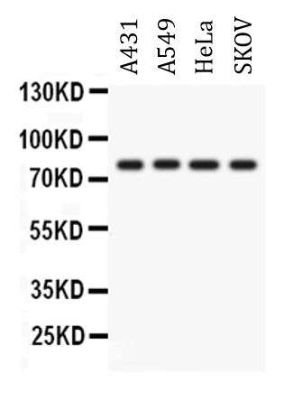
ARG42739 anti-PKC eta antibody WB image
Western blot: 50 µg of sample under reducing conditions. A431, A549, HeLa and SKOV whole cell lysates stained with ARG42739 anti-PKC eta antibody at 0.5 µg/ml dilution, overnight at 4°C.
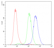
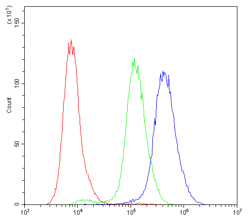
ARG42739 anti-PKC eta antibody FACS image
Flow Cytometry: K562 cells were blocked with 10% normal goat serum and then stained with ARG42739 anti-PKC eta antibody (blue) at 1 µg/10^6 cells for 30 min at 20°C, followed by incubation with DyLight®488 labelled secondary antibody. Isotype control antibody (green) was Rabbit IgG (1 µg/10^6 cells) used under the same conditions. Unlabelled sample (red) was also used as a control.
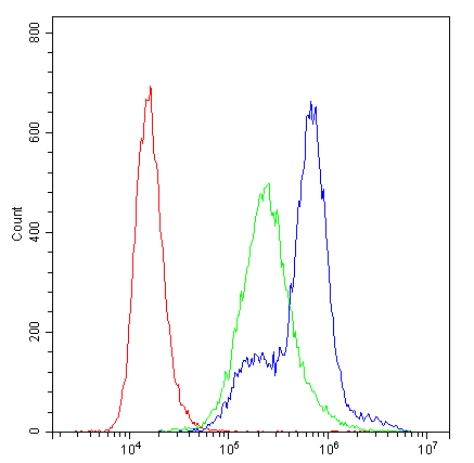
ARG42739 anti-PKC eta antibody FACS image
Flow Cytometry: MCF7 cells were blocked with 10% normal goat serum and then stained with ARG42739 anti-PKC eta antibody (blue) at 1 µg/10^6 cells for 30 min at 20°C, followed by incubation with DyLight®488 labelled secondary antibody. Isotype control antibody (green) was Rabbit IgG (1 µg/10^6 cells) used under the same conditions. Unlabelled sample (red) was also used as a control.
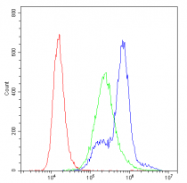
ARG42739 anti-PKC eta antibody FACS image
Flow Cytometry: A431 cells were blocked with 10% normal goat serum and then stained with ARG42739 anti-PKC eta antibody (blue) at 1 µg/10^6 cells for 30 min at 20°C, followed by incubation with DyLight®488 labelled secondary antibody. Isotype control antibody (green) was Rabbit IgG (1 µg/10^6 cells) used under the same conditions. Unlabelled sample (red) was also used as a control.


 New Products


