


ATP1A1/ATP1A2/ATP1A3 Mouse Monoclonal Antibody (H-3)
Key features and details
- Reactivity:Human, Mouse, Rat
- Application:WB, IP, IF, IHC(P) , ELISA
- Host:Mouse
- Clonality:Monoclonal
- lsotype:IgG2bκ
- Target Name:ATP1A1/ATP1A2/ATP1A3
-
Brand:

CAT.NO. : ARA1233
RMB Please choose
RMB Please choose
Size:
Trail, Bulk size or Custom requests Please contact us
*产品价格可能会有所调整,请以品牌方官网实时更新的价格为准,以确保准确性。
 Product Details
Background
The ubiquitously expressed sodium/potassium - ATPase (Na⁺/K⁺ - ATPase) exists as an oligomeric plasma membrane complex that couples the hydrolysis of one molecule of ATP to the importation of three Na⁺ ions and two K⁺ ions against their respective electrochemical gradients. As a member of the P - type family of ion motives, Na⁺/K⁺ - ATPase plays a critical role in maintaining cellular volume, resting membrane potential and Na⁺ - coupled solute transport. Multiple isoforms of three subunits, α, β and γ, comprise the Na⁺/K⁺ - ATPase oligomer. The α subunit contains the binding sites for ATP and the cations; the glycosylated β subunit ensures correct folding and membrane insertion of the α subunits. The small γ subunit co - localizes with the α subunit in nephron segments, where it increases the affinity of Na⁺/K⁺ - ATPase for ATP. The β subunit, but not the γ subunit, is essential for normal activity of Na⁺/K⁺ - ATPase.
Application
Na⁺/K⁺ - ATPase α (H - 3) is recommended for detection of Na⁺/K⁺ - ATPase α1, 2 and 3 of mouse, rat and human origin by Western Blotting (starting dilution 1:2000, dilution range 1:2000 - 1:10000), immunoprecipitation [1 - 2 µg per 100 - 500 µg of total protein (1 ml of cell lysate)], immunofluorescence (starting dilution 1:50, dilution range 1:50 - 1:500), immunohistochemistry (including paraffin - embedded sections) (starting dilution 1:50, dilution range 1:50 - 1:500) and solid phase ELISA (starting dilution 1:30, dilution range 1:30 - 1:3000).
Data

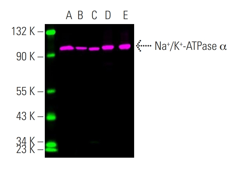
Na⁺/K⁺ - ATPase α (H - 3) Alexa Fluor® 546. Direct fluorescent western blot analysis of Na⁺/K⁺ - ATPase α expression in PC - 12 (A), Hep G2 (B) and MDCK (C) whole cell lysates and human kidney (D) and human brain (E) tissue extracts.


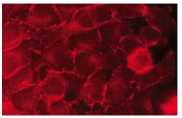
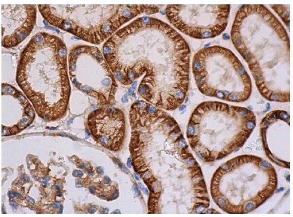
Na⁺/K⁺ - ATPase α (H - 3). Immunofluorescence staining of methanol - fixed HeLa cells showing membrane localization (above). Immunoperoxidase staining of formalin fixed, paraffin - embedded human kidney tissue showing cytoplasmic staining of cells in glomeruli in membrane and cytoplasmic staining of cells in tubules (below).
New Products


