


BAX Rabbit Monoclonal Antibody(ARA865)

Key features and details
- Reactivity:Human
- Application:IHC
- Host:Rabbit
- Clonality:Monoclonal
- Target:BAX
-
Brand:

CAT.NO. : ARA6691
RMB Please choose
RMB Please choose
*产品价格可能会有所调整,请以品牌方官网实时更新的价格为准,以确保准确性。
 Product Details
Background
Apoptosis regulator BAX, also known as bcl-2-like protein 4, is a protein that in humans is encoded by the BAX gene. BAX is a member of the Bcl-2 gene family. BCL2 family members form hetero- or homodimers and act as anti- or pro-apoptotic regulators that are involved in a wide variety of cellular activities. This protein forms a heterodimer with BCL2, and functions as an apoptotic activator. This protein is reported to interact with, and increase the opening of, the mitochondrial voltage-dependent anion channel (VDAC), which leads to the loss in membrane potential and the release of cytochrome c. The expression of this gene is regulated by the tumor suppressor P53 and has been shown to be involved in P53-mediated apoptosis. In healthy mammalian cells, the majority of BAX is found in the cytosol, but upon initiation of apoptotic signaling, Bax undergoes a conformational shift. Upon induction of apoptosis, BAX becomes organelle membrane-associated, and in particular, mitochondrial membrane associated. BAX is believed to interact with, and induce the opening of the mitochondrial voltage-dependent anion channel, VDAC. Alternatively, growing evidence also suggests that activated BAX and/or Bak form an oligomeric pore, MAC in the MOM (mitochondrial outer membrane). This results in the release of cytochrome c and other pro-apoptotic factors from the mitochondria, often referred to as mitochondrial outer membrane permeabilization, leading to activation of caspases. This defines a direct role for BAX in mitochondrial outer membrane permeabilization. BAX activation is stimulated by various abiotic factors, including heat, hydrogen peroxide, low or high pH, and mitochondrial membrane remodeling. In addition, it can become activated by binding BCL-2, as well as non-BCL-2 proteins such as p53 and Bif-1. Conversely, BAX can become inactivated by interacting with VDAC2, Pin1, and IBRDC2.
Application
|
Application |
Dilution Ratio |
|
IHC-P |
1:200 |
Overview
|
Antibody Type |
Recombinant Rabbit monoclonal Antibody |
|
Immunogen |
Synthetic peptide within Human Bax aa 50-150 (internal sequence) |
|
Species Reactivity |
Human |
|
Validated Applications |
IHC-P |
|
Molecular Weight |
Predicted band size: 21 kDa |
|
Positive Control |
Human breast tissue, human cervical carcinoma tissue, human kidney tissue |
|
Conjugation |
unconjugated |
|
Form |
Liquid |
|
Concentration |
1μg/ul |
|
Storage Buffer |
PBS (pH7.4), 0.1% BSA, 40% Glycerol, Preservative: 0.05% Sodium Azide |
|
Isotype |
IgG |
|
Purification Method |
Protein A affinity purified |
Data

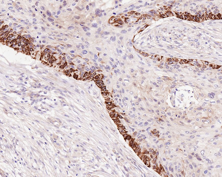
Immunohistochemical analysis of paraffin-embedded human cervical carcinoma tissue with BAX Rabbit Monoclonal Antibody(ARA865)
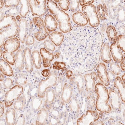
Immunohistochemical analysis of paraffin-embedded human kidney tissue with BAX Rabbit Monoclonal Antibody(ARA865)
Storage
Store at 4°C short term. For long term storage, store at -20°C, avoiding freeze/thaw cycles.
Research Use Only
For Research Use Only. Not for use in diagnostic procedures.
New Products


