ERK1 Rabbit Monoclonal Antibody(ARA780)


Key features and details
- Reactivity:Human, Mouse, Rat
- Application:WB, IHC-P, IP, FC, ICC/IF
- Host:Rabbit
- Clonality:Monoclonal
- Target:ERK1
-
Brand:

CAT.NO. : ARA6567
RMB Please choose
RMB Please choose
Size:
Trail, Bulk size or Custom requests Please contact us
*产品价格可能会有所调整,请以品牌方官网实时更新的价格为准,以确保准确性。
 Product Details
Background
ERK1 and ERK2 belongs to the protein kinase superfamily. It is involved in both the initiation and regulation of meiosis, mitosis, and postmitotic functions in differentiated cells by phosphorylating a number of transcription factors such as ELK-1. ERK1/2 catalyzed the reaction: ATP + a protein = ADP + a phosphoprotein. It is activated by tyrosine phosphorylation in response to insulin and NGF.
Application
|
Application |
Dilution Ratio |
|
WB |
1:2,000 - 1:10,000 |
|
IHC-P |
1:100 - 1:200 |
|
IP |
1:10 - 1:50 |
|
FC |
1:200 - 1:1,000 |
|
ICC/IF |
1:50 - 1:200 |
Overview
|
Predicted Molecular Weight |
43kDa |
|
Species Cross-reactivity |
Human, Mouse, Rat (Species cross-reactivity determined by WB) |
|
Validated Applications |
WB, IHC-P, IF/ICC, FC, IP |
|
Purity |
ProA affinity purified IgG |
|
Form |
Liquid |
|
Swissprot ID |
P27361 |
|
Immunogen |
A synthetic peptide corresponding to the N-term of ERK1 was used as an immunogen. |
|
Storage Buffer |
PBS 59%, Sodium azide 0.01%, Glycerol 40%, BSA 0.05%. |
Data
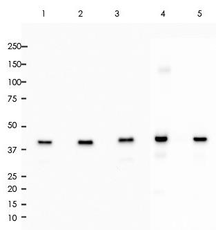
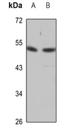
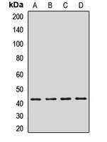
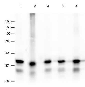
All lanes: Anti-ERK1 antibody at 1:2,000 dilution
Predicted MW: 43 kDa
Observed MW: 44 kDa
Lane 1: Hela
Lane 2: 293T
Lane 3: Raw264.7
Lane 4: A431
Lane 5: A375
Lysate at 10 μg per lane
2nd Ab: GAR HRP(H+L) 1:5,000
Exposure: 50s

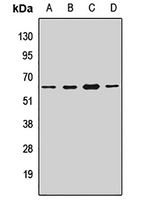
All lanes: Anti-ERK1 antibody at 1:2,000 dilution
Predicted MW: 43 kDa
Observed MW: 44 kDa
Lane 1: Mu Brain
Lane 2: Mu Kidney
Lane 3: Mu Liver
Lane 4: Rat Brain
Lane 5: Rat Kidney
Lysate at 10 μg per lane
2nd Ab: GAR HRP(H+L) 1:5,000
Exposure: 20s
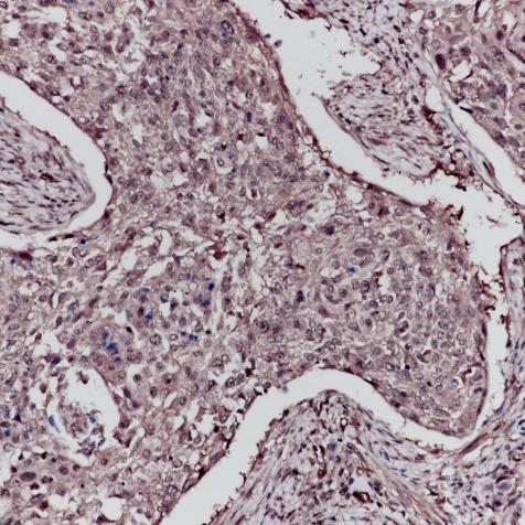
Immunohistochemistry (Formalin/PFA-fixed paraffin paraffin-embedded sections) analysis Cervix cancer tissue labelling ERK1 with ERK1 Rabbit Monoclonal Antibody(ARA780)

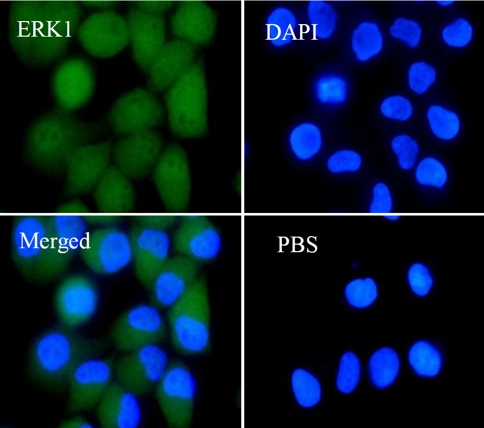
ERK1 Rabbit Monoclonal Antibody(ARA780) staining ERK1 in Hela cells by IF/ICC (immunofluorescence/immunocytochemistry).
Cells were fixed with paraformaldehyde, permeabilized with 0.1% Triton X-100 and blocked with 10% goat serum for half an hour at room temperature.
Samples were incubated with primary antibody (1:50) at 4°C.
An Alexa Fluor® 488-conjugated Goat Anti-Rabbit IgG polyclonal was used as the secondary antibody (1:500).
DAPI (blue) was used as the nuclear counter stain.
Control: PBS and secondary antibody, An Alexa Fluor® 488-conjugated Goat Anti-Rabbit IgG (1:500).
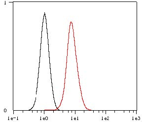
Overlay histogram showing Hela cells stained with ERK1 Rabbit Monoclonal Antibody(ARA780) (Red).
The cells were fixed with 4% paraformaldehyde (10 min) and then permeabilized with 0.1% TritonX-100 for 15 min.
The cells were then incubated in the antibody (ERK1 Rabbit Monoclonal Antibody(ARA780), 1:1,000 dilution) in 1x PBS/1% BSA for 30 min at room temperature.
The secondary antibody used was a Goat Anti-Rabbit Alexa Fluor® 488 (IgG H+L) at 1:2,000 dilution for 20 min at room temperature.
Unlabelled sample (Black) was used as a control.
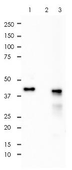
ERK1 was immunoprecipitated from 0.4mg of A375 whole cell lysate with ERK1 Rabbit Monoclonal Antibody(ARA780) at 1:50 dilution.
2nd Ab:
GAR HRP for IP 1:500
Lane 1: ERK1 Rabbit Monoclonal Antibody(ARA780) IP in A375 whole cell lysate
Lane 2: PBS instead of ERK1 Rabbit Monoclonal Antibody(ARA780) in A375 whole cell lysate
Lane 3: A375 whole cell lysate, 10 μg (input)
Exposure: 120s
Storage
Store at 4°C short term. For long term storage, store at -20°C, avoiding freeze/thaw cycles.
Research Use Only
For Research Use Only. Not for use in diagnostic procedures.
New Products