


GLS Rabbit Polyclonal Antibody


Key features and details
- Reactivity:Human, Mouse, Rat
- Application:WB, IHC, IF/ICC
- Host:Rabbit
- Clonality:Polyclonal
- Target:GLS
-
Brand:

 Product Details
Glutaminase is a phosphate-activated amidohydrolase that catalyzes the hydrolysis of glutamine to glutamate and ammonia. It is primarily expressed in the brain and kidney and plays an essential role in generating energy for metabolism, synthesizing neurotransmitter glutamate, and maintaing acid-based balance in the kidney. There are alternate splicing results in multiple transcript variants in the Glutaminase gene.
|
Application |
Dilution Ratio |
|
WB |
1:500 - 1:1000 |
|
IHC |
1:50 - 1:200 |
|
IF/ICC |
1:50 - 1:200 |
|
Immunogen |
Recombinant full length protein of human GLS |
|
Purification Method |
The antibody was purified by immunogen affinity chromatography |
|
Clonality |
Polyclonal |
|
Form |
Liquid in 0.42% Potassium phosphate, 0.87% Sodium chloride, pH 7.3, 30% glycerol, and 0.01% sodium azide |
|
Gene Symbol |
GLS |
|
Alternative Names |
GLS1; KIAA0838; Glutaminase kidney isoform mitochondrial; GLS; K-glutaminase; L-glutamine amidohydrolase |
|
Gene ID (Human) |
2744 |
|
Gene ID (Mouse) |
14660 |
|
Protein ID (Human) |
O94925 |
|
Protein ID (Mouse) |
D3Z7P3 |
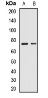
Western blot analysis of GLS expression in SW620 (A), mouse lung (B) whole cell lysates. (Predicted band size: 17; 65; 73 kD; Observed band size: 73 kD)
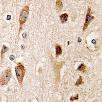
Immunohistochemical analysis of GLS staining in human brain formalin fixed paraffin embedded tissue section. The section was pre-treated using heat mediated antigen retrieval with sodium citrate buffer (pH 6.0). The section was then incubated with the antibody at room temperature and detected using an HRP conjugated compact polymer system. DAB was used as the chromogen. The section was then counterstained with haematoxylin and mounted with DPX.

Immunofluorescent analysis of GLS staining in NIH3T3 cells. Formalin-fixed cells were permeabilized with 0.1% Triton X-100 in TBS for 5-10 minutes and blocked with 3% BSA-PBS for 30 minutes at room temperature. Cells were probed with the primary antibody in 3% BSA-PBS and incubated overnight at 4 °C in a hidified chamber. Cells were washed with PBST and incubated with a AF488-conjugated secondary antibody (green) in PBS at room temperature in the dark. Phalloidin - AF594 was used to stain Actin filaments (red). DAPI was used to stain the cell nuclei (blue).
New Products


