


Human IL-3 ELISA Kit
 Protocol Booklet

Key features and details
- Specification:
- Sensitivity:
- Standard Curve Range:
- Standard Curve Gradient:
- Number of Incubations:
- Sample Volume:
- Assay type:
- Operation Duration:
-
Brand:

CAT.NO. : AEH0050
RMB Please choose
RMB Please choose
Size:
Trail, Bulk size or Custom requests Please contact us
*产品价格可能会有所调整,请以品牌方官网实时更新的价格为准,以确保准确性。
 Product Details
Background
Interleukin 3 is a pleiotropic factor produced primarily by activated T cells that can stimulate the proliferation and differentiation of pluripotent hematopoietic stem cells as well as various lineage committed progenitors. In addition, IL-3 also affects the functional activity of mature mast cells, basophils, eosinophils and macrophages. Because of its multiple functions and targets, it was originally studied under different names, including mast cell growth factor P-cell stimulating factor, burst promoting activity, multi-colony stimulating factor, thy-1 inducing factor and WEHI-3 growth factor. In addition to activated T cells, other cell types such as human thymic epithelial cells, activated mouse mast cells, mouse keratinocytes and neurons/astrocytes can also produce IL-3. At the amino acid sequence level, mature human and mouse IL-3 share only 29% sequence identity. Consistent with this lack of homology, IL-3 activity is highly species-specific and human IL-3 does not show activity on mouse cells.
IL-3 exerts its biological activities through binding to specific cell surface receptors. The high affinity receptor responsible for IL-3 signaling is composed of alpha and beta subunits. The IL-3 R alpha is a member of the cytokine receptor super family and binds IL-3 with low affinity. Two distinct beta subunits, AIC2A ( beta IL-3) and AIC2B ( beta c) are present in mouse cells. beta IL-3 also binds IL-3 with low affinity and forms a high affinity receptor with the alpha subunit. The beta c subunits does not bind any cytokine but forms functional high affinity receptors with the alpha subunit of the IL-3, IL-5 and GM-CSF receptors. Receptors for IL-3 are present on bone marrow progenitors, macrophages, mast cells, eosinophils, megakaryocytes, basophils and various myeloid leukemic cells.
IL-3 exerts its biological activities through binding to specific cell surface receptors. The high affinity receptor responsible for IL-3 signaling is composed of alpha and beta subunits. The IL-3 R alpha is a member of the cytokine receptor super family and binds IL-3 with low affinity. Two distinct beta subunits, AIC2A ( beta IL-3) and AIC2B ( beta c) are present in mouse cells. beta IL-3 also binds IL-3 with low affinity and forms a high affinity receptor with the alpha subunit. The beta c subunits does not bind any cytokine but forms functional high affinity receptors with the alpha subunit of the IL-3, IL-5 and GM-CSF receptors. Receptors for IL-3 are present on bone marrow progenitors, macrophages, mast cells, eosinophils, megakaryocytes, basophils and various myeloid leukemic cells.
Typical data
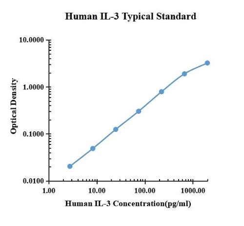
| pg/ml | O.D. | Average | Corrected | |
| 0.00 | 0.0157 | 0.0159 | 0.0158 | |
| 40.96 | 0.0347 | 0.0360 | 0.0354 | 0.0240 |
| 102.40 | 0.0625 | 0.0650 | 0.0638 | 0.0488 |
| 256.00 | 0.1395 | 0.1391 | 0.1393 | 0.1243 |
| 640.00 | 0.3246 | 0.3103 | 0.3175 | 0.3025 |
| 1600.00 | 0.8032 | 0.8016 | 0.8024 | 0.7874 |
| 4000.00 | 1.9100 | 1.9030 | 1.9065 | 1.8915 |
| 10000.00 | 3.2770 | 3.1920 | 3.2345 | 3.2195 |
Precision
| Intra-assay Precision | Inter-assay Precision | |||||
| Sample Number | S1 | S2 | S3 | S1 | S2 | S3 |
| 22 | 22 | 22 | 6 | 6 | 6 | |
| Average(pg/ml) | 244.6 | 1239.7 | 3546.7 | 191.8 | 849.4 | 2573 |
| Standard Deviation | 21.5 | 110.5 | 309.9 | 7.3 | 31.2 | 141.2 |
| Coefficient of Variation(%) | 8.8 | 8.9 | 8.7 | 3.8 | 3.7 | 5.5 |
Intra-assay Precision (Precision within an assay) Three samples of known concentration were tested twenty times on one plate to assess intra-assay precision.
Inter-assay Precision (Precision between assays) Three samples of known concentration were tested six times on one plate to assess intra-assay precision.
Spike Recovery
The spike recovery was evaluated by spiking 3 levels of human IL-3 into health human serum sample. The un-spiked serum was used as blank in this experiment.
The recovery ranged from 165% to 197% with an overall mean recovery of 184%.
The recovery ranged from 165% to 197% with an overall mean recovery of 184%.
Sample Values
| Sample Matrix | Sample Evaluated | Range (pg/ml) | Detectable (%) | Mean of Detectable (pg/ml) |
| Serum | 30 | n.d.-40.96 | 37 | 20.6 |
Serum/Plasma – Thirty samples from apparently healthy volunteers were evaluated for the presence of IL-12/IL-23 p40 in this assay. No medical histories were available for the donors.
New Products


