


Integrin β1/ITGB1 Mouse Monoclonal Antibody (4B7R)
 Datasheet


Key features and details
- Reactivity:Human, Mouse, Rat
- Application:IF, IHC(P), FCM,ELISA
- Host:Mouse
- Clonality:Monoclonal
- lsotype:IgG1κ
- Target Name:Integrin β1/ITGB1
-
Brand:

CAT.NO. : ARA1147
RMB Please choose
RMB Please choose
Size:
Trail, Bulk size or Custom requests Please contact us
*产品价格可能会有所调整,请以品牌方官网实时更新的价格为准,以确保准确性。
 Product Details
Background
Integrins are heterodimers composed of noncovalently associated transmembrane α and β subunits. The 16 α and 8 β subunits heterodimerize to produce more than 20 different receptors. Most integrin receptors bind ligands that are components of the extracellular matrix, including Fibronectin, collagen and Vitronectin. Certain integrins can also bind to soluble ligands such as Fibrinogen, or to counterreceptors on adjacent cells such as the intracellular adhesion molecules (ICAMs), leading to aggregation of cells. Ligands serve to cross - link or cluster integrins by binding to adjacent integrin receptors; both receptor clustering and ligand occupancy are necessary for the activation of integrin - mediated responses. In addition to mediating cell adhesion and cytoskeletal organization, integrins function as signaling receptors. Signals transduced by integrins play a role in many biological processes, including cell growth, differentiation, migration and apoptosis.
Application
Integrin β1 (4B7R) is recommended for detection of Integrin β1 of mouse, rat and human origin by immunofluorescence (starting dilution 1:50, dilution range 1:50 - 1:500), immunohistochemistry (including paraffin - embedded sections) (starting dilution 1:50, dilution range 1:50 - 1:500), flow cytometry (1 µg per 1 x 10⁶ cells) and solid phase ELISA (starting dilution 1:30, dilution range 1:30 - 1:3000).
Data

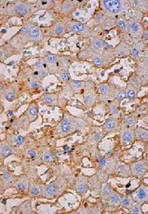
Integrin β1 (4B7R). Immunofluorescence staining of methanol - fixed HUV - EC - C cells showing membrane and cytoplasmic localization (Left). Immunoperoxidase staining of formalin fixed, paraffin - embedded human liver tissue showing membrane staining of hepatocytes (Right).
New Products


