PIP5K Rabbit Polyclonal Antibody

Key features and details
- Reactivity:Human, Rat
- Application:WB, IF/IC
- Host:Rabbit
- Clonality:Polyclonal
- Target:PIP5K
-
Brand:

CAT.NO. : ARA6824
RMB Please choose
RMB Please choose
*产品价格可能会有所调整,请以品牌方官网实时更新的价格为准,以确保准确性。
 Product Details
Background
Anti-PIP5K antibodies enable researchers to detect and measure the PIP5K antigen in biological samples. This target is a reported synonym of the PIKFYVE gene, which encodes phosphoinositide kinase, FYVE-type zinc finger containing. This protein is known to function in intracellular signal transduction, among other biological roles. The human version of PIP5K has a canonical amino acid length of 2098 residues and a protein mass of 237.1 kilodaltons, although 4 isoforms have been identified. It is reported to be localized in the cytoplasmic vesicles and membrane of cells. Western Blot is the most common application for the PIP5K antibodies listed below. ELISA, Immunofluorescence, and Immunohistochemistry are also common applications.
Application
|
Application |
Dilution Ratio |
|
WB |
1:500 - 1:2,000 |
|
IF/IC |
1:50 - 1:100 |
Overview
|
Immunogen |
KLH-conjugated synthetic peptide encompassing a sequence within the N-term region of human PIP5K. The exact sequence is proprietary |
|
Purification Method |
The antibody was purified by immunogen affinity chromatography |
|
Clonality |
Polyclonal |
|
Form |
Liquid in 0.42% Potassium phosphate, 0.87% Sodium chloride, pH 7.3, 30% glycerol, and 0.01% sodium azide |
|
Gene Name |
PIKFYVE |
|
Synonyms |
KIAA0981; PIP5K3; 1-phosphatidylinositol 3-phosphate 5-kinase; Phosphatidylinositol 3-phosphate 5-kinase; FYVE finger-containing phosphoinositide kinase; PIKfyve; Phosphatidylinositol 3-phosphate 5-kinase type III; PIPKin-III; Type III PIP kinase |
|
Gene ID (Human) |
200576 |
|
Protein ID (Human) |
Q9Y2Y7 |
Data
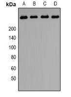
Western blot analysis of PIP5K expression in A549 (A), HepG2 (B), PC3 (C)rat testis (D) whole cell lysates. (Predicted band size: 237 kD; Observed band size: 262 kD)

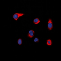
Immunofluorescent analysis of PIP5K staining in HepG2 cells. Formalin-fixed cells were permeabilized with 0.1% Triton X-100 in TBS for 5-10 minutes and blocked with 3% BSA-PBS for 30 minutes at room temperature. Cells were probed with the primary antibody in 3% BSA-PBS and incubated overnight at 4 °C in a hidified chamber. Cells were washed with PBST and incubated with a DyLight 594-conjugated secondary antibody (red) in PBS at room temperature in the dark. DAPI was used to stain the cell nuclei (blue).
Storage
Store at 4°C short term. For long term storage, store at -20°C, avoiding freeze/thaw cycles.
Research Use Only
For Research Use Only. Not for use in diagnostic procedures.
New Products