


T-cell infiltration Antibody Duo


Key features and details
- 产品描述:
- 靶点名称:
- 別名:
-
Brand:

 Product Details
| 货号 | 内含物名称 | 宿主克隆性 | 反应 | 应用 | 包装 |
|---|---|---|---|---|---|
| ARG65859 | anti-CD3 epsilon antibody [SQab1713] | Rabbit mAb | Hu | FACS, ICC/IF, IHC-P, IP, WB | 50 μl |
| ARG65860 | anti-CD4 antibody [SQab1714] | Rabbit mAb | Hu | FACS, IHC-P, IP, WB | 50 μl |
| 产品描述 | T-cells have the ability to leave the bloodstream and migrate into and attack tumor cells. Evidences show that enhanced T-cell infiltration in tumor tissue result to increased survival. T-cell infiltration is critical for examining the effect of cancer immunotherapy.arigo’s T-cell Infiltration Antibody Duo offers two quality antibodies against CD3 and CD4. It is the best solution for labeling infiltrating T-cells in tissue. |
|---|---|
| 靶点名称 | T-cell infiltration |
| 別名 | T-cell infiltration antibody; CD3 antibody; CD4 antibody |
| 存放说明 | For continuous use, store undiluted antibody at 2-8°C for up to a week. For long-term storage, aliquot and store at -20°C or below. Storage in frost free freezers is not recommended. Avoid repeated freeze/thaw cycles. Suggest spin the vial prior to opening. The antibody solution should be gently mixed before use. |
|---|---|
| 注意事项 | For laboratory research only, not for drug, diagnostic or other use. |
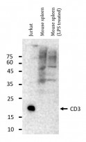
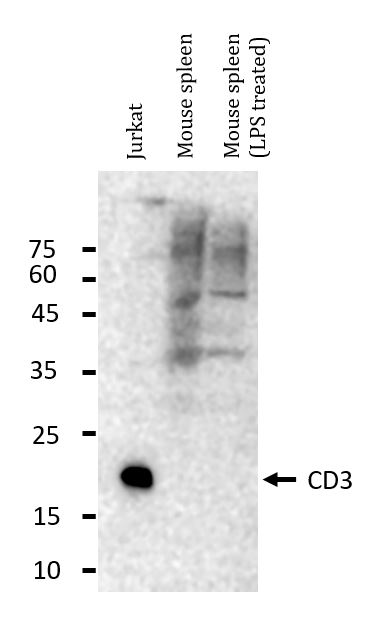
ARG65859 anti-CD3 epsilon antibody [SQab1713] WB image (Customer's Feedback)
Western blot: 20 µg of Jurkat and Mouse spleen (untreated or treated with LPS) lysates stained with ARG65859 anti-CD3 epsilon antibody [SQab1713] at 1:1000 dilution, overnight at 4°C.

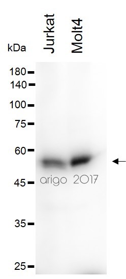
ARG65860 anti-CD4 antibody [SQab1714] WB image
Western blot: 30 µg of Jurkat and Molt4 cell lysates stained with ARG65860 anti-CD4 antibody [SQab1714] at 1:500 dilution.

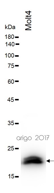
ARG65859 anti-CD3 epsilon antibody [SQab1713] WB image (Customer's Feedback)
Western blot: 30 µg of Molt4 cell lysate stained with ARG65859 anti-CD3 epsilon antibody [SQab1713] at 1:500 dilution.

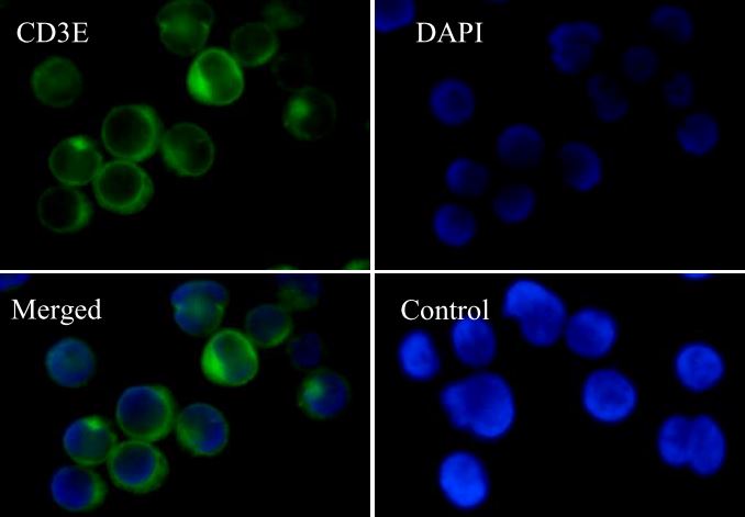
ARG65859 anti-CD3 epsilon antibody [SQab1713] ICC/IF image
Immunofluorescence: Jurkat cells were fixed with 4% paraformaldehyde for 30 min at RT, permeabilized with 0.1% Triton X-100 for 10 min at RT then blocked with 10% Goat serum for half an hour at room temperature. Samples were stained with ARG65859 anti-CD3 epsilon antibody [SQab1713] (green) at 1:50 and 4°C. DAPI (blue) was used as the nuclear counter stain. Control: PBS and secondary antibody.
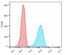
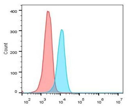
ARG65859 anti-CD3 epsilon antibody [SQab1713] FACS image
Flow Cytometry: Jurkat cells were fixed with 4% paraformaldehyde for 10 min. The cells were then stained with ARG65859 anti-CD3 epsilon antibody [SQab1713] (blue) at 1:1000 dilution in 1x PBS/1% BSA for 30 min at room temperture, followed by Alexa Fluor® 488 labelled secondary antibody. Unlabelled sample (red) was used as a control.

ARG65860 anti-CD4 antibody [SQab1714] FACS image
Flow Cytometry: Jurkat cells were fixed with 4% paraformaldehyde for 10 min. The cells were then stained with ARG65860 anti-CD4 antibody [SQab1714] (blue) at 1:50 dilution in 1x PBS/1% BSA for 30 min at room temperture, followed by Alexa Fluor® 488 labelled secondary antibody. Unlabelled sample (red) was used as a control.

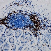
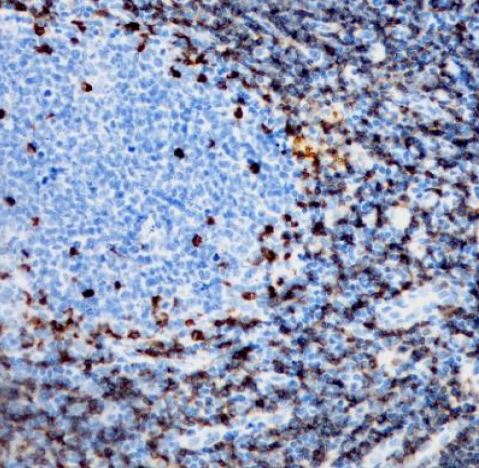
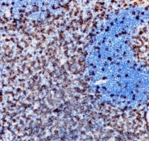
ARG65859 anti-CD3 epsilon antibody [SQab1713] IHC-P image
Immunohistochemistry: Formalin/PFA-fixed and paraffin-embedded sections of Human tonsil tissue stained with ARG65859 anti-CD3 epsilon antibody [SQab1713] at 1:200 dilution. Antigen Retrieval: Boil tissue section in Tris/EDTA buffer (pH 9.0).
ARG65860 anti-CD4 antibody [SQab1714] IHC-P image
Immunohistochemistry: Formalin/PFA-fixed and paraffin-embedded sections of Human tonsil tissue stained with ARG65860 anti-CD4 antibody [SQab1714] at 1:2000 dilution. Antigen Retrieval: Boil tissue section in Tris/EDTA buffer (pH 9.0).
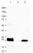
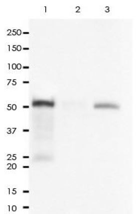
ARG65859 anti-CD3 epsilon antibody [SQab1713] IP image
Immunoprecipitation: 0.4 mg of Molt-4 whole cell lysate was immunoprecipitated (1:15 dilution) and stained with ARG65859 anti-CD3 epsilon antibody [SQab1713].
Lane 1: Immunoprecipitation in Molt-4 whole cell lysate
Lane 2: Rabbit IgG instead of Primary Ab in Molt-4 whole cell lysate
Lane 3: Molt-4 whole cell lysate, 10 µg (input)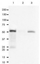
ARG65860 anti-CD4 antibody [SQab1714] IP image
Immunoprecipitation: 0.4 mg of Molt-4 whole cell lysate was immunoprecipitated (1:50 dilution) and stained with ARG65860 anti-CD4 antibody [SQab1714].
Lane 1: Immunoprecipitation in Molt-4 whole cell lysate
Lane 2: Rabbit IgG instead of Primary Ab in Molt-4 whole cell lysate
Lane 3: Molt-4 whole cell lysate, 10 µg (input)ARG65859 anti-CD3 epsilon antibody [SQab1713] IHC-P image
Immunohistochemistry: Formalin/PFA-fixed and paraffin-embedded sections of Human colon tissue stained with ARG65859 anti-CD3 epsilon antibody [SQab1713] at 1:200 dilution. Antigen Retrieval: Boil tissue section in Tris/EDTA buffer (pH 9.0).


 New Products


