Adipophilin Rabbit Polyclonal Antibody


Key features and details
- Reactivity:Human, Mouse, Rat
- Application:WB, IHC, IF/ICC
- Host:Rabbit
- Clonality:Polyclonal
- Target:Adipophilin
-
Brand:

CAT.NO. : ARA6868
RMB Please choose
RMB Please choose
*产品价格可能会有所调整,请以品牌方官网实时更新的价格为准,以确保准确性。
 Product Details
Background
Milk lipid globules from humans, cows and rats contain a protein identified as adipocyte differentiation-related protein (ADFP). It is associated with the globule surface membrane material. This protein, previously believed to be specific to adipocytes, is a major constituent of the globule surface and is present in a detergent-insoluble complex that contains butyrophilin and xanthine oxidase. ADFP (Adipophilin) is found in a wide range of cultured cell lines, including fibroblasts, endothelial and epithelial cells. In tissues, however, expression of adipophilin is restricted to specific cell types, such as lactating mammary epithelial cells, adrenal cortex cells, Sertoli and Leydig cells of the male reproductive system, and steatosis or fatty change hepatocytes in alcoholic liver cirrhosis. ADFP may be a possible new marker for the identification of specialized differentiated cells containing lipid droplets and for diseases associated with fat-accumulating cells.
Application
|
Application |
Dilution Ratio |
|
WB |
1:500 - 1:1000 |
|
IHC |
1:50 - 1:200 |
|
IF/ICC |
1:50 - 1:200 |
Overview
|
Immunogen |
Recombinant full length protein of human Adipophilin |
|
Purification Method |
The antibody was purified by immunogen affinity chromatography. |
|
Clonality |
Polyclonal |
|
Product Form |
Liquid in 0.42% Potassium phosphate, 0.87% Sodium chloride, pH 7.3, 30% glycerol, and 0.01% sodium azide. |
|
Gene Name |
PLIN2 |
|
Alternative Names |
ADFP; Perilipin-2; Adipophilin; Adipose differentiation-related protein; ADRP |
|
Human Gene ID |
123 |
|
Mouse Gene ID |
11520 |
|
Human Protein ID |
Q99541 |
|
Mouse Protein ID |
P43883 |
Data
-
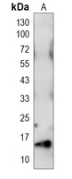
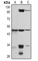
Western blot analysis of Adipophilin expression in HepG2 (A), mouse brain (B) , rat heart (C) whole cell lysates. (Predicted band size: 48 kD; Observed band size: 48 kD)
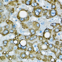
Immunohistochemical analysis of Adipophilin staining in human liver formalin fixed paraffin embedded tissue section. The section was pre-treated using heat mediated antigen retrieval with sodium citrate buffer (pH 6.0). The section was then incubated with the antibody at room temperature and detected using an HRP conjugated compact polymer system. DAB was used as the chromogen. The section was then counterstained with haematoxylin and mounted with DPX.

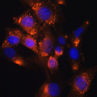
Immunofluorescent analysis of Adipophilin staining in NIH3T3 cells. Formalin-fixed cells were permeabilized with 0.1% Triton X-100 in TBS for 5-10 minutes and blocked with 3% BSA-PBS for 30 minutes at room temperature. Cells were probed with the primary antibody in 3% BSA-PBS and incubated overnight at 4 °C in a humidified chamber. Cells were washed with PBST and incubated with a AF594-conjugated secondary antibody (red) in PBS at room temperature in the dark. DAPI was used to stain the cell nuclei (blue).
Storage
Store at 4°C short term. For long term storage, store at -20°C, avoiding freeze/thaw cycles.
Research Use Only
For Research Use Only. Not for use in diagnostic procedures.
New Products