


anti-c-Cbl antibody

Key features and details
- 产品描述:
- 反应物种:
- 应用:
- 宿主:
- 克隆:
- 同位型:
- 靶点名称:
- 抗原物种:
- 抗原:
-
Brand:

 Product Details
| 产品描述 | Rabbit Polyclonal antibody recognizes c-Cbl |
|---|---|
| 反应物种 | Hu, Ms, Rat |
| 应用 | FACS, ICC/IF, IHC-P, WB |
| 宿主 | Rabbit |
| 克隆 | Polyclonal |
| 同位型 | IgG |
| 靶点名称 | c-Cbl |
| 抗原物种 | Human |
| 抗原 | Recombinant protein corresponding to A556-T906 of Human c-Cbl. |
| 偶联标记 | Un-conjugated |
| 別名 | Signal transduction protein CBL; C-CBL; EC 6.3.2.-; FRA11B; Casitas B-lineage lymphoma proto-oncogene; Proto-oncogene c-Cbl; RNF55; CBL2; E3 ubiquitin-protein ligase CBL; RING finger protein 55; NSLL |
| 应用建议 |
| ||||||||||
|---|---|---|---|---|---|---|---|---|---|---|---|
| 应用说明 | * The dilutions indicate recommended starting dilutions and the optimal dilutions or concentrations should be determined by the scientist. | ||||||||||
| 实际分子量 | ~ 110 kDa |
| 形式 | Liquid |
|---|---|
| 纯化 | Affinity purification with immunogen. |
| 缓冲液 | 0.2% Na2HPO4, 0.9% NaCl, 0.05% Sodium azide and 5% BSA. |
| 抗菌剂 | 0.05% Sodium azide |
| 稳定剂 | 5% BSA |
| 浓度 | 0.5 mg/ml |
| 存放说明 | For continuous use, store undiluted antibody at 2-8°C for up to a week. For long-term storage, aliquot and store at -20°C or below. Storage in frost free freezers is not recommended. Avoid repeated freeze/thaw cycles. Suggest spin the vial prior to opening. The antibody solution should be gently mixed before use. |
| 注意事项 | For laboratory research only, not for drug, diagnostic or other use. |
| 数据库连接 | |
|---|---|
| 基因名称 | CBL |
| 全名 | Cbl proto-oncogene, E3 ubiquitin protein ligase |
| 背景介绍 | This gene is a proto-oncogene that encodes a RING finger E3 ubiquitin ligase. The encoded protein is one of the enzymes required for targeting substrates for degradation by the proteasome. This protein mediates the transfer of ubiquitin from ubiquitin conjugating enzymes (E2) to specific substrates. This protein also contains an N-terminal phosphotyrosine binding domain that allows it to interact with numerous tyrosine-phosphorylated substrates and target them for proteasome degradation. As such it functions as a negative regulator of many signal transduction pathways. This gene has been found to be mutated or translocated in many cancers including acute myeloid leukaemia, and expansion of CGG repeats in the 5' UTR has been associated with Jacobsen syndrome. Mutations in this gene are also the cause of Noonan syndrome-like disorder. [provided by RefSeq, Jul 2016] |
| 生物功能 | Adapter protein that functions as a negative regulator of many signaling pathways that are triggered by activation of cell surface receptors. Acts as an E3 ubiquitin-protein ligase, which accepts ubiquitin from specific E2 ubiquitin-conjugating enzymes, and then transfers it to substrates promoting their degradation by the proteasome. Recognizes activated receptor tyrosine kinases, including KIT, FLT1, FGFR1, FGFR2, PDGFRA, PDGFRB, EGFR, CSF1R, EPHA8 and KDR and terminates signaling. Recognizes membrane-bound HCK, SRC and other kinases of the SRC family and mediates their ubiquitination and degradation. Participates in signal transduction in hematopoietic cells. Plays an important role in the regulation of osteoblast differentiation and apoptosis. Essential for osteoclastic bone resorption. The 'Tyr-731' phosphorylated form induces the activation and recruitment of phosphatidylinositol 3-kinase to the cell membrane in a signaling pathway that is critical for osteoclast function. May be functionally coupled with the E2 ubiquitin-protein ligase UB2D3. In association with CBLB, required for proper feedback inhibition of ciliary platelet-derived growth factor receptor-alpha (PDGFRA) signaling pathway via ubiquitination and internalization of PDGFRA (By similarity). [UniProt] |
| 细胞定位 | Cytoplasm. Cell membrane. Note=Colocalizes with FGFR2 in lipid rafts at the cell membrane. [UniProt] |
| 预测分子量 | 100 kDa |
| 翻译后修饰 | Phosphorylated on tyrosine residues by ALK, EGFR, SYK, FYN and ZAP70 (By similarity). Phosphorylated on tyrosine residues in response to FLT1 and KIT signaling. Phosphorylated on tyrosine residues by INSR and FGR. Phosphorylated on several tyrosine residues by constitutively activated FGFR3. Not phosphorylated at Tyr-731 by FGFR3. Phosphorylated on tyrosine residues by activated CSF1R, PDGFRA and PDGFRB. Phosphorylated on tyrosine residues by HCK. Ubiquitinated, leading to its degradation via the proteasome. [UniProt] |

ARG42688 anti-c-Cbl antibody ICC/IF image
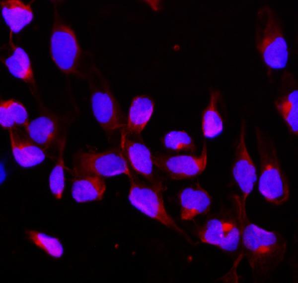
Immunofluorescence: U2OS cells were blocked with 10% goat serum and then stained with ARG42688 anti-c-Cbl antibody (red) at 2 µg/ml dilution, overnight at 4°C. DAPI (blue) for nuclear staining.
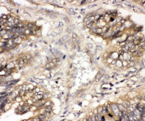
ARG42688 anti-c-Cbl antibody IHC-P image
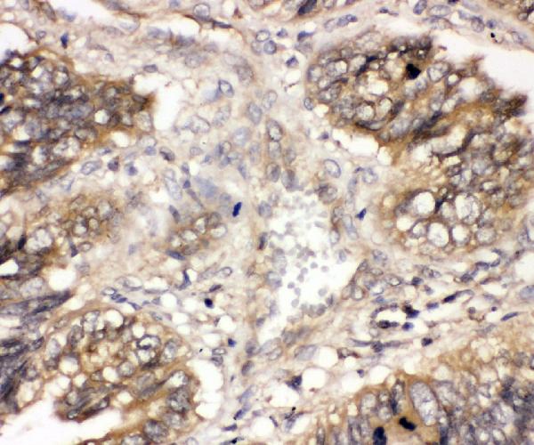
Immunohistochemistry: Paraffin-embedded Human intestinal cancer tissue. Antigen Retrieval: Heat mediation was performed in EDTA buffer (pH 8.0). The tissue section was blocked with 10% goat serum. The tissue section was then stained with ARG42688 anti-c-Cbl antibody at 1 µg/ml dilution, overnight at 4°C.

ARG42688 anti-c-Cbl antibody WB image
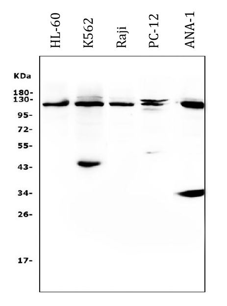
Western blot: 50 µg of samples under reducing conditions. HL-60, K562, Raji, PC-12 and ANA-1 whole cell lysates stained with ARG42688 anti-c-Cbl antibody at 0.5 µg/ml dilution, overnight at 4°C.
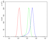
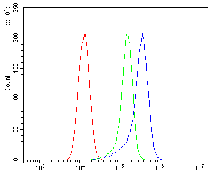
ARG42688 anti-c-Cbl antibody FACS image
Flow Cytometry: A549 cells were blocked with 10% normal goat serum and then stained with ARG42688 anti-c-Cbl antibody (blue) at 1 µg/10^6 cells for 30 min at 20°C, followed by incubation with DyLight®488 labelled secondary antibody. Isotype control antibody (green) was rabbit IgG (1 µg/10^6 cells) used under the same conditions. Unlabelled sample (red) was also used as a control.
ARG42688 anti-c-Cbl antibody IHC-P image
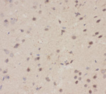
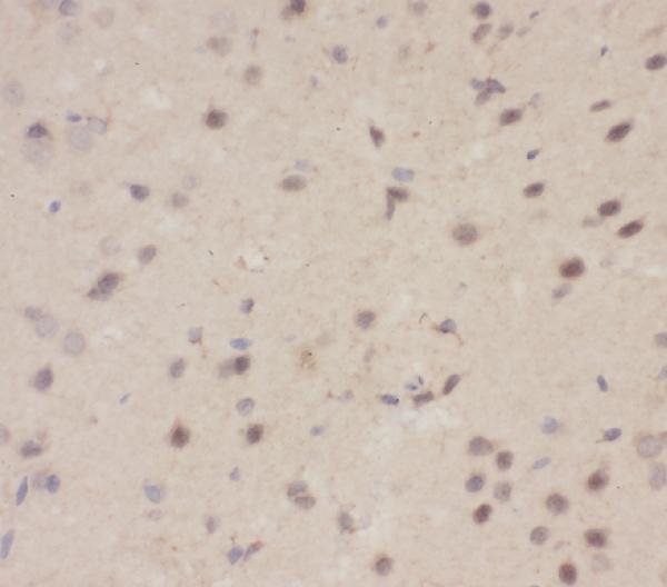
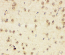
Immunohistochemistry: Paraffin-embedded Mouse brain tissue. Antigen Retrieval: Heat mediation was performed in EDTA buffer (pH 8.0). The tissue section was blocked with 10% goat serum. The tissue section was then stained with ARG42688 anti-c-Cbl antibody at 1 µg/ml dilution, overnight at 4°C.
ARG42688 anti-c-Cbl antibody IHC-P image
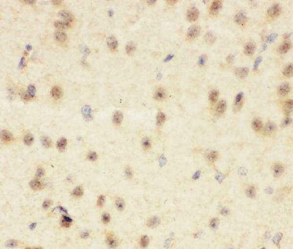
Immunohistochemistry: Paraffin-embedded Rat brain tissue. Antigen Retrieval: Heat mediation was performed in EDTA buffer (pH 8.0). The tissue section was blocked with 10% goat serum. The tissue section was then stained with ARG42688 anti-c-Cbl antibody at 1 µg/ml dilution, overnight at 4°C.


 New Products


