


anti-Calbindin antibody [5A9]
![anti-Calbindin antibody [5A9]](/upload/image/products/ARG10702_IHC_2_210_205.jpg)
![anti-Calbindin antibody [5A9]](/upload/image/products/ARG10702_IHC-Fr_190729.jpg)
![anti-Calbindin antibody [5A9]](/upload/image/products/ARG10702_IHC_1.jpg)
![anti-Calbindin antibody [5A9]](/upload/image/products/ARG10702_WB_1.jpg)
![anti-Calbindin antibody [5A9]](/upload/image/products/ARG10702_WB_190729.jpg)
![anti-Calbindin antibody [5A9]](/upload/image/products/ARG10702_IHC_2.jpg)
Key features and details
- 产品描述:
- 反应物种:
- 预测物种:
- 应用:
- 宿主:
- 克隆:
- 克隆号:
- 同位型:
- 靶点名称:
-
Brand:

 Product Details
| 产品描述 | Mouse Monoclonal antibody [5A9] recognizes Calbindin |
|---|---|
| 反应物种 | Hu, Ms, Rat, Cow, Hrs, Pig |
| 预测物种 | Chk |
| 应用 | ICC/IF, IHC-Fr, WB |
| 宿主 | Mouse |
| 克隆 | Monoclonal |
| 克隆号 | 5A9 |
| 同位型 | IgG2a |
| 靶点名称 | Calbindin |
| 抗原物种 | Human |
| 抗原 | Full length recombinant Human protein. |
| 偶联标记 | Un-conjugated |
| 別名 | Vitamin D-dependent calcium-binding protein, avian-type; Calbindin; CALB; Calbindin D28; D-28K |
| 应用建议 |
| ||||||||
|---|---|---|---|---|---|---|---|---|---|
| 应用说明 | * The dilutions indicate recommended starting dilutions and the optimal dilutions or concentrations should be determined by the scientist. |
| 形式 | Liquid |
|---|---|
| 纯化 | Affinity purification. |
| 缓冲液 | PBS and 50% Glycerol. |
| 稳定剂 | 50% Glycerol |
| 浓度 | 1 mg/ml |
| 存放说明 | For continuous use, store undiluted antibody at 2-8°C for up to a week. For long-term storage, aliquot and store at -20°C. Storage in frost free freezers is not recommended. Avoid repeated freeze/thaw cycles. Suggest spin the vial prior to opening. The antibody solution should be gently mixed before use. |
| 注意事项 | For laboratory research only, not for drug, diagnostic or other use. |
| 数据库连接 | |
|---|---|
| 基因名称 | CALB1 |
| 全名 | calbindin 1, 28kDa |
| 背景介绍 | The protein encoded by this gene is a member of the calcium-binding protein superfamily that includes calmodulin and troponin C. Originally described as a 27 kDa protein, it is now known to be a 28 kDa protein. It contains four active calcium-binding domains, and has two modified domains that are thought to have lost their calcium binding capability. This protein is thought to buffer entry of calcium upon stimulation of glutamate receptors. Depletion of this protein was noted in patients with Huntington disease. [provided by RefSeq, Jan 2015] |
| 生物功能 | Buffers cytosolic calcium. May stimulate a membrane Ca(2+)-ATPase and a 3',5'-cyclic nucleotide phosphodiesterase. [UniProt] |
| 产品亮点 | Related products: Calbindin antibodies; Anti-Mouse IgG secondary antibodies; Related news: Viral-like capsids, new trans-synaptic mRNA transport mechanism |
| 预测分子量 | 30 kDa |
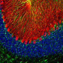
ARG10702 anti-Calbindin antibody [5A9] IHC-Fr image
Immunohistochemistry: Frozen section of Rat brain cerebellum stained with ARG10702 anti-Calbindin antibody [5A9] (red) at 1:2000 dilution and costained with ARG52312 anti-GFAP antibody (green) at 1:5000 dilution. DAPI (blue) for nuclear staining. (Sample preparation: Following transcardial perfusion with 4% paraformaldehyde, brain was post fixed for 24 hours, cut to 45 µM, and free-floating sections were stained with the above antibodies.)
The Clone 5A9 calbindin antibody prominently labels the dendrites and perikarya of Purkinje cells in the molecular layer of the cerebellum. The GFAP antibody stains the processes of Bergmann glia in the molecular layer and astroglia in the granular and white matter layers of cerebellum.
ARG10702 anti-Calbindin antibody [5A9] IHC-Fr image
Immunohistochemistry: Frozen sections of adult Mouse cortex were stained with ARG10702 anti-Calbindin antibody [5A9] (red), and co-stained with our rabbit polyclonal anti-Fox3 / NeuN antibody (green). Calbindin is expressed in a subset of interneurons in the cortex. Fox3 / NeuN expresses in most neurons; as a result, cells positive for calbindin appear to be yellow. The Inset is a high magnification image of the boxed area. Blue is DAPI nucleus staining that labels DNA.
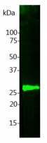
ARG10702 anti-Calbindin antibody [5A9] WB image
Western blot: Cow cerebellum lysate were stained with ARG10702 anti-Calbindin antibody [5A9].
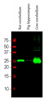
ARG10702 anti-Calbindin antibody [5A9] WB image
Western blot: Rat cerebellum, Pig hippocampus and Cow cerebellum lysates stained with ARG10702 anti-Calbindin antibody [5A9] (green) at 1:5000 dilution.
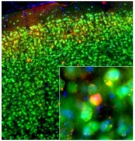
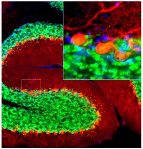
ARG10702 anti-Calbindin antibody [5A9] IHC-Fr image
Immunohistochemistry: Frozen sections of adult Mouse cerebellum were stained with ARG10702 anti-Calbindin antibody [5A9] (red), and co-stained with our rabbit polyclonal anti-Fox3 / NeuN antibody (green). Calbindin is prominently expressed in the dendrites of Purkinje cells in the cerebellum molecular layer. Fox3 / NeuN expresses in most neurons; as a result, cells positive for calbindin appear to be yellow. The Inset is a high magnification image of the boxed area. Blue is DAPI nucleus staining that labels DNA.


 New Products


