


anti-CD107a / LAMP1 antibody [5H6]
![anti-CD107a / LAMP1 antibody [5H6]](/upload/image/products/ARG52327_WB_1_191002_210_205.jpg)
![anti-CD107a / LAMP1 antibody [5H6]](/upload/image/products/ARG52327_WB_Sheet1_Rev1_160425.jpg)
![anti-CD107a / LAMP1 antibody [5H6]](/upload/image/products/ARG52327_IF_1_191002.jpg)
![anti-CD107a / LAMP1 antibody [5H6]](/upload/image/products/ARG52327_WB_1_191002.jpg)
Key features and details
- 产品描述:
- 反应物种:
- 不反应物种:
- 应用:
- 宿主:
- 克隆:
- 克隆号:
- 同位型:
- 靶点名称:
-
Brand:

 Product Details
| 产品描述 | Mouse Monoclonal antibody [5H6] recognizes CD107a / LAMP1 |
|---|---|
| 反应物种 | Hu, Bov |
| 不反应物种 | Ms, Rat |
| 应用 | ICC/IF, WB |
| 宿主 | Mouse |
| 克隆 | Monoclonal |
| 克隆号 | 5H6 |
| 同位型 | IgG1 |
| 靶点名称 | CD107a / LAMP1 |
| 抗原物种 | Human |
| 抗原 | Recombinant full length human LAMP1 expressed in and purified from E. coli |
| 偶联标记 | Un-conjugated |
| 別名 | LGP120; CD107a; LAMPA; CD antigen CD107a; Lysosome-associated membrane glycoprotein 1; CD107 antigen-like family member A; LAMP-1; Lysosome-associated membrane protein 1 |
| 应用建议 |
| ||||||
|---|---|---|---|---|---|---|---|
| 应用说明 | Specific for the ~100k LAMP1 protein * The dilutions indicate recommended starting dilutions and the optimal dilutions or concentrations should be determined by the scientist. |
| 形式 | Liquid |
|---|---|
| 纯化 | Affinity Purified |
| 缓冲液 | PBS and 10 mM Sodium azide |
| 抗菌剂 | 10 mM Sodium azide |
| 存放说明 | For continuous use, store undiluted antibody at 2-8°C for up to a week. For long-term storage, aliquot and store at -20°C or below. Storage in frost free freezers is not recommended. Avoid repeated freeze/thaw cycles. Suggest spin the vial prior to opening. The antibody solution should be gently mixed before use. |
| 注意事项 | For laboratory research only, not for drug, diagnostic or other use. |
| 数据库连接 | Swiss-port # P11279 Human Lysosome-associated membrane glycoprotein 1 Swiss-port # Q05204 Bovine Lysosome-associated membrane glycoprotein 1 |
|---|---|
| 基因名称 | LAMP1 |
| 全名 | lysosomal-associated membrane protein 1 |
| 背景介绍 | Lysosomal Associated Membrane Protein1 (LAMP1) is a protein that is localized primarily in lysosomes but may also be present on late endosomes and the plasma membrane. LAMP1 antibodies are therefore widely used as lysosome markers. It has recently been suggested that lysosomes are activated in microglia in the progression of multiple system atrophy (MSA) and thus play a key role in its pathology (Makioka et al., 2012). |
| 产品亮点 | Related Antibody Duos and Panels: ARG30310 Endosome, Lysosome, Peroxisome Marker Antibody Panel (Catalase, Caveolin1, Clathrin heavy chain, LAMP1) Related products: LAMP1 antibodies; LAMP1 Duos / Panels; Anti-Mouse IgG secondary antibodies; Related poster download: Organelle Markers & Loading Control |
| 研究领域 | Cancer antibody; Cell Death antibody; Controls and Markers antibody; Developmental Biology antibody; Metabolism antibody; Neuroscience antibody; Signaling Transduction antibody; Lysosome Marker antibody |
| 预测分子量 | 45 kDa |
| 翻译后修饰 | O- and N-glycosylated; some of the 18 N-linked glycans are polylactosaminoglycans. The glycosylation of N-76 is essential for Lassa virus entry into cells. |
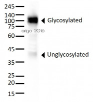
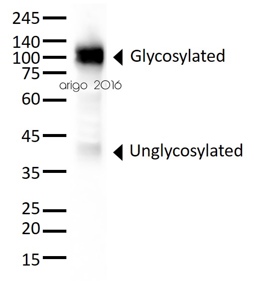
ARG52327 anti-CD107a / LAMP1 antibody [5H6] WB image
Western blot: 30 µg of HeLa cell lysate stained with ARG52327 anti-CD107a / LAMP1 antibody [5H6] at 1:5000 dilution.

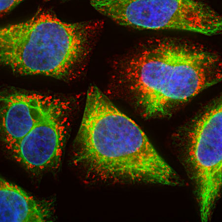
ARG52327 anti-CD107a / LAMP1 antibody [5H6] ICC/IF image
Immunofluorescence: HeLa cells were treated with 50 µM of chloroquine, an inhibitor of autophagy, for 16 hours prior to staining. Cells stained with ARG52327 anti-CD107a / LAMP1 antibody [5H6] (red) at 1:500 dilution, and costained with ARG52468 anti-Vimentin antibody (green) at 1:10000 dilution. DAPI (blue) for nuclear staining.
Clone 5H6 reveals vesicular staining of LAMP1 protein accumulated in swollen lysosomes, while the Vimentin antibody specifically labels the intermediate filament network in these cells.
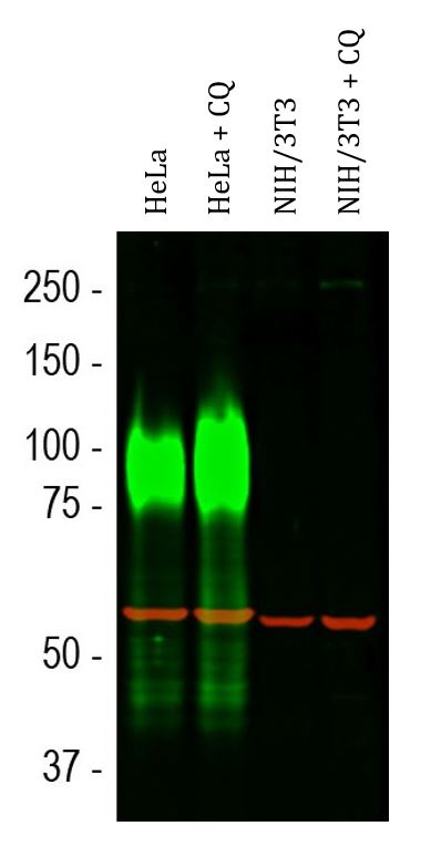
ARG52327 anti-CD107a / LAMP1 antibody [5H6] WB image
Western blot: Cells were untreated or treated with 50 µM of chloroquine (CQ), an inhibitor of autophagy, for 24 hours. HeLa, HeLa + CQ, NIH/3T3 and NIH/3T3 + CQ (left to right) cell lysates stained with ARG52327 anti-CD107a / LAMP1 antibody [5H6] (green) at 1:10000 dilution. The smeared band between 75-120 kDa corresponds to variably glycosylated forms of the LAMP1 protein detected only in the Human cells, this antibody does not recognize the rodent LAMP1 homologue.
The same blot was stained with ARG10757 anti-Hsp 60 antibody (red) at 1:20000 dilution.
 New Products


