


anti-CD279 / PD-1 antibody [SQab1732]
![anti-CD279 / PD-1 antibody [SQab1732]](/upload/image/products/ARG66243_IF_293.jpg)
![anti-CD279 / PD-1 antibody [SQab1732]](/upload/image/products/ARG66243_FACS_293.jpg)
![anti-CD279 / PD-1 antibody [SQab1732]](/upload/image/products/ARG66243_IHC_1.jpg)
![anti-CD279 / PD-1 antibody [SQab1732]](/upload/image/products/ARG66243_IP_293.jpg)
Key features and details
- 产品描述:
- 反应物种:
- 应用:
- 宿主:
- 克隆:
- 克隆号:
- 同位型:
- 靶点名称:
- 抗原物种:
-
Brand:

 Product Details
| 产品描述 | Recombinant Rabbit Monoclonal antibody [SQab1732] recognizes CD279 / PD-1 |
|---|---|
| 反应物种 | Hu |
| 应用 | FACS, ICC/IF, IHC-P, IP |
| 宿主 | Rabbit |
| 克隆 | Monoclonal |
| 克隆号 | SQab1732 |
| 同位型 | IgG |
| 靶点名称 | CD279 / PD-1 |
| 抗原物种 | Human |
| 抗原 | Recombinant full length protein of Human PD‐1. The immunogen contains the specific extracellular domain of Human PD‐1 (Leu25 ‐ Gln167). |
| 偶联标记 | Un-conjugated |
| 別名 | hPD-l; CD279; PD-1; Protein PD-1; CD antigen CD279; PD1; hSLE1; SLEB2; Programmed cell death protein 1; hPD-1 |
| 应用建议 |
| ||||||||||
|---|---|---|---|---|---|---|---|---|---|---|---|
| 应用说明 | IHC-P: Antigen Retrieval: Boil tissue section in Tris/EDTA buffer (pH 9.0). * The dilutions indicate recommended starting dilutions and the optimal dilutions or concentrations should be determined by the scientist. |
| 形式 | Liquid |
|---|---|
| 纯化 | Purification with Protein A. |
| 缓冲液 | PBS, 0.01% Sodium azide, 40% Glycerol and 0.05% BSA. |
| 抗菌剂 | 0.01% Sodium azide |
| 稳定剂 | 40% Glycerol and 0.05% BSA |
| 存放说明 | For continuous use, store undiluted antibody at 2-8°C for up to a week. For long-term storage, aliquot and store at -20°C. Storage in frost free freezers is not recommended. Avoid repeated freeze/thaw cycles. Suggest spin the vial prior to opening. The antibody solution should be gently mixed before use. |
| 注意事项 | For laboratory research only, not for drug, diagnostic or other use. |
| 数据库连接 | |
|---|---|
| 基因名称 | PDCD1 |
| 全名 | programmed cell death 1 |
| 背景介绍 | CD279 / PD-1 is a cell surface membrane protein of the immunoglobulin superfamily. This protein is expressed in pro-B-cells and is thought to play a role in their differentiation. In mice, expression of this gene is induced in the thymus when anti-CD3 antibodies are injected and large numbers of thymocytes undergo apoptosis. Mice deficient for this gene bred on a BALB/c background developed dilated cardiomyopathy and died from congestive heart failure. These studies suggest that this gene product may also be important in T cell function and contribute to the prevention of autoimmune diseases. [provided by RefSeq, Jul 2008] |
| 生物功能 | CD279 / PD-1 is an inhibitory receptor on antigen activated T-cells. It plays a critical role in induction and maintenance of immune tolerance to self (PubMed:21276005). Delivers inhibitory signals upon binding to ligands CD274/PDCD1L1 and CD273/PDCD1LG2 (PubMed:21276005). Following T-cell receptor (TCR) engagement, PDCD1 associates with CD3-TCR in the immunological synapse and directly inhibits T-cell activation. Suppresses T-cell activation through the recruitment of PTPN11/SHP-2: following ligand-binding, PDCD1 is phosphorylated within the ITSM motif, leading to the recruitment of the protein tyrosine phosphatase PTPN11/SHP-2 that mediates dephosphorylation of key TCR proximal signaling molecules, such as ZAP70, PRKCQ/PKCtheta and CD247/CD3zeta. The PDCD1-mediated inhibitory pathway is exploited by tumors to attenuate anti-tumor immunity and escape destruction by the immune system, thereby facilitating tumor survival (PubMed:28951311). The interaction with CD274/PDCD1L1 inhibits cytotoxic T lymphocytes (CTLs) effector function (PubMed:28951311). The blockage of the PDCD1-mediated pathway results in the reversal of the exhausted T-cell phenotype and the normalization of the anti-tumor response, providing a rationale for cancer immunotherapy (PubMed:22658127, PubMed:25034862, PubMed:25399552). [UniProt] |
| 产品亮点 | Related products: PD-1 antibodies; PD-1 ELISA Kits; PD-1 Duos / Panels; Anti-Rabbit IgG secondary antibodies; Related news: Cancer Pathology Markers (SQ clones) The best solution for PD-1/PD-L1 research Examining CTL/NK-mediated cytotoxicity by ELISA |
| 预测分子量 | 32 kDa |
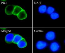
ARG66243 anti-CD279 / PD-1 antibody [SQab1732] ICC/IF image
Immunofluorescence: 293 cells transfected with PD-1 gene, fixed with 4% paraformaldehyde for 30 min at RT, permeabilized with 0.1% Triton X-100 for 10 min at RT then blocked with 10% Goat serum for half an hour at room temperature. Samples were stained with ARG66243 anti-CD279 / PD-1 antibody [SQab1732] (green) at 1:10000 and 4°C. DAPI (blue) was used as the nuclear counter stain. Control: PBS and secondary antibody.

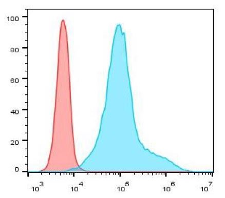
ARG66243 anti-CD279 / PD-1 antibody [SQab1732] FACS image
Flow Cytometry: 293 cells transfected with PD-1 gene. The cells were fixed with 4% paraformaldehyde (10 min) and then permeabilized with 0.1% TritonX-100 for 15 min. The cells were then stained with ARG66243 anti-CD279 / PD-1 antibody [SQab1732] (blue) at 1:200 dilution in 1x PBS/1% BSA for 30 min at room temperature, followed by Alexa Fluor® 488 labelled secondary antibody. Unlabelled sample (red) was used as a control.
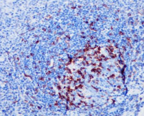
ARG66243 anti-CD279 / PD-1 antibody [SQab1732] IHC-P image
Immunohistochemistry: Formalin-fixed and paraffin-embedded Human lung tonsil tissue stained with ARG66243 anti-CD279 / PD-1 antibody [SQab1732] at 1:200. Antigen Retrieval: Boil tissue section in Tris/EDTA buffer (pH 9.0).
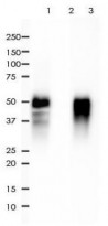
ARG66243 anti-CD279 / PD-1 antibody [SQab1732] IP image
Immunoprecipitation: 0.2 mg of 293 whole cell lysate (transfected with PD-1 gene) was immunoprecipitated (1:50 dilution) by ARG66243 anti-CD279 / PD-1 antibody [SQab1732] and then stained with anti-CD279 / PD-1 antibody.
Lane 1: Immunoprecipitation in 293 whole cell lysate (transfected with PD-1 gene)
Lane 2: PBS instead of Primary Ab in 293 whole cell lysate (transfected with PD-1 gene)
Lane 3: 293 whole cell lysate (transfected with PD-1 gene), 2 µg (input)


Comparative transcriptomic analysis reveals differences in gene expression and regulatory pathways between nonacral and acral melanoma in Asian individuals
IHC-P / Human

 New Products


