anti-CD3 epsilon antibody [SQab1713]
![anti-CD3 epsilon antibody [SQab1713]](/upload/image/products/ARG65859_IHC_Hu_colon_210_205.jpg)
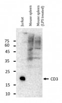
![anti-CD3 epsilon antibody [SQab1713]](/upload/image/products/ARG65859_WB_210603_Sheet_Rev.jpg)
![anti-CD3 epsilon antibody [SQab1713]](/upload/image/products/ARG65859_WB_Sheet_Rev_Molt4_170609.jpg)
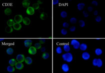
![anti-CD3 epsilon antibody [SQab1713]](/upload/image/products/ARG65859_IF_Jurkat.jpg)
![anti-CD3 epsilon antibody [SQab1713]](/upload/image/products/ARG65859_FACS_Jurkat.jpg)
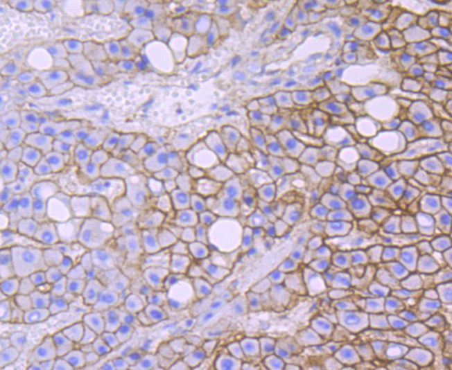
![anti-CD3 epsilon antibody [SQab1713]](/upload/image/products/ARG65859_IHC_Hu_tonsil.jpg)
![anti-CD3 epsilon antibody [SQab1713]](/upload/image/products/ARG65859_IP_1.jpg)
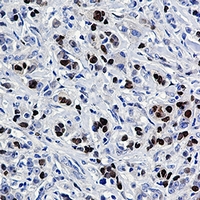
![anti-CD3 epsilon antibody [SQab1713]](/upload/image/products/ARG65859_IHC_Hu_colon.jpg)
Key features and details
- 产品描述:
- 反应物种:
- 应用:
- 特异性:
- 宿主:
- 克隆:
- 克隆号:
- 同位型:
- 靶点名称:
-
Brand:

 Product Details
| 产品描述 | Recombinant Rabbit Monoclonal antibody [SQab1713] recognizes CD3 |
|---|---|
| 反应物种 | Hu |
| 应用 | FACS, ICC/IF, IHC-P, IP, WB |
| 特异性 | This antibody has been tested not reacting to mouse spleen lysates by WB. |
| 宿主 | Rabbit |
| 克隆 | Monoclonal |
| 克隆号 | SQab1713 |
| 同位型 | IgG |
| 靶点名称 | CD3 epsilon |
| 抗原物种 | Human |
| 抗原 | Synthetic peptide around the N-terminus of Human CD3 epsilon. |
| 偶联标记 | Un-conjugated |
| 別名 | CD3E; CD3 Epsilon Subunit Of T-Cell Receptor Complex; T-Cell Surface Glycoprotein CD3 Epsilon Chain; CD3e Antigen, Epsilon Polypeptide (TiT3 Complex); T-Cell Surface Antigen T3/Leu-4 Epsilon Chain; CD3e Molecule, Epsilon (CD3-TCR Complex); CD3-Epsilon; CD3epsilon |
| 应用建议 |
| ||||||||||||
|---|---|---|---|---|---|---|---|---|---|---|---|---|---|
| 应用说明 | IHC-P: Antigen Retrieval: Boil tissue section in Tris/EDTA buffer (pH 9.0). * The dilutions indicate recommended starting dilutions and the optimal dilutions or concentrations should be determined by the scientist. |
| 形式 | Liquid |
|---|---|
| 纯化 | Purification with Protein A. |
| 缓冲液 | PBS, 0.01% Sodium azide, 40% Glycerol and 0.05% BSA. |
| 抗菌剂 | 0.01% Sodium azide |
| 稳定剂 | 40% Glycerol and 0.05% BSA |
| 存放说明 | For continuous use, store undiluted antibody at 2-8°C for up to a week. For long-term storage, aliquot and store at -20°C. Storage in frost free freezers is not recommended. Avoid repeated freeze/thaw cycles. Suggest spin the vial prior to opening. The antibody solution should be gently mixed before use. |
| 注意事项 | For laboratory research only, not for drug, diagnostic or other use. |
| 数据库连接 | Swiss-port # P07766 Human T-cell surface glycoprotein CD3 epsilon chain |
|---|---|
| 基因名称 | CD3E |
| 全名 | CD3 Epsilon Subunit Of T-Cell Receptor Complex |
| 背景介绍 | The protein encoded by this gene is the CD3-epsilon polypeptide, which together with CD3-gamma, -delta and -zeta, and the T-cell receptor alpha/beta and gamma/delta heterodimers, forms the T-cell receptor-CD3 complex. This complex plays an important role in coupling antigen recognition to several intracellular signal-transduction pathways. The genes encoding the epsilon, gamma and delta polypeptides are located in the same cluster on chromosome 11. The epsilon polypeptide plays an essential role in T-cell development. Defects in this gene cause immunodeficiency. This gene has also been linked to a susceptibility to type I diabetes in women. |
| 生物功能 | Part of the TCR-CD3 complex present on T-lymphocyte cell surface that plays an essential role in adaptive immune response. When antigen presenting cells (APCs) activate T-cell receptor (TCR), TCR-mediated signals are transmitted across the cell membrane by the CD3 chains CD3D, CD3E, CD3G and CD3Z. All CD3 chains contain immunoreceptor tyrosine-based activation motifs (ITAMs) in their cytoplasmic domain. Upon TCR engagement, these motifs become phosphorylated by Src family protein tyrosine kinases LCK and FYN, resulting in the activation of downstream signaling pathways. |
| 细胞定位 | Cell membrane, Membrane |
| 产品亮点 | Related Antibody Duos and Panels: ARG30302 T-cell infiltration Antibody Duo ARG30325 Inflammatory Cell Antibody Panel ARG30334 Tumor-infiltrating Lymphocyte Antibody Panel Related products: CD3 antibodies; CD3 ELISA Kits; CD3 Duos / Panels; CD3 recombinant proteins; Anti-Rabbit IgG secondary antibodies; Related news: More effective cocktail therapy for cancer immune evasion Cholesterol, the weakness of anaplastic large cell lymphoma (ALCL) New antibody panels and duos for Tumor immune microenvironment Tumor-Infiltrating Lymphocytes (TILs) Exploring Antiviral Immune Response |
| 研究领域 | Cancer antibody; Developmental Biology antibody; Immune System antibody; Lymphocyte Marker antibody; Inflammatory Cell Marker antibody; T-cell Marker antibody; T-cell infiltration Study antibody; Tumor-infiltrating Lymphocyte Study antibody |
| 预测分子量 | 23 kDa |
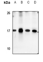
ARG65859 anti-CD3 epsilon antibody [SQab1713] WB image (Customer's Feedback)
Western blot: 20 µg of Jurkat and Mouse spleen (untreated or treated with LPS) lysates stained with ARG65859 anti-CD3 epsilon antibody [SQab1713] at 1:1000 dilution, overnight at 4°C.

ARG65859 anti-CD3 epsilon antibody [SQab1713] WB image (Customer's Feedback)
Western blot: 30 µg of Molt4 cell lysate stained with ARG65859 anti-CD3 epsilon antibody [SQab1713] at 1:500 dilution.
ARG65859 anti-CD3 epsilon antibody [SQab1713] ICC/IF image
Immunofluorescence: Jurkat cells were fixed with 4% paraformaldehyde for 30 min at RT, permeabilized with 0.1% Triton X-100 for 10 min at RT then blocked with 10% Goat serum for half an hour at room temperature. Samples were stained with ARG65859 anti-CD3 epsilon antibody [SQab1713] (green) at 1:50 and 4°C. DAPI (blue) was used as the nuclear counter stain. Control: PBS and secondary antibody.
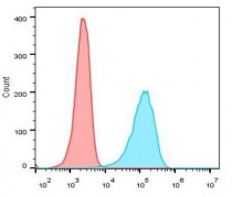
ARG65859 anti-CD3 epsilon antibody [SQab1713] FACS image
Flow Cytometry: Jurkat cells were fixed with 4% paraformaldehyde for 10 min. The cells were then stained with ARG65859 anti-CD3 epsilon antibody [SQab1713] (blue) at 1:1000 dilution in 1x PBS/1% BSA for 30 min at room temperture, followed by Alexa Fluor® 488 labelled secondary antibody. Unlabelled sample (red) was used as a control.
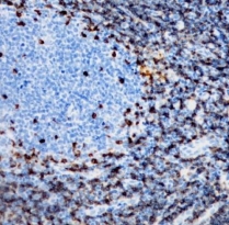
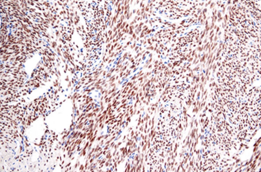
ARG65859 anti-CD3 epsilon antibody [SQab1713] IHC-P image
Immunohistochemistry: Formalin/PFA-fixed and paraffin-embedded sections of Human tonsil tissue stained with ARG65859 anti-CD3 epsilon antibody [SQab1713] at 1:200 dilution. Antigen Retrieval: Boil tissue section in Tris/EDTA buffer (pH 9.0).
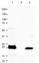
ARG65859 anti-CD3 epsilon antibody [SQab1713] IP image
Immunoprecipitation: 0.4 mg of Molt-4 whole cell lysate was immunoprecipitated (1:15 dilution) and stained with ARG65859 anti-CD3 epsilon antibody [SQab1713].
Lane 1: Immunoprecipitation in Molt-4 whole cell lysate
Lane 2: Rabbit IgG instead of Primary Ab in Molt-4 whole cell lysate
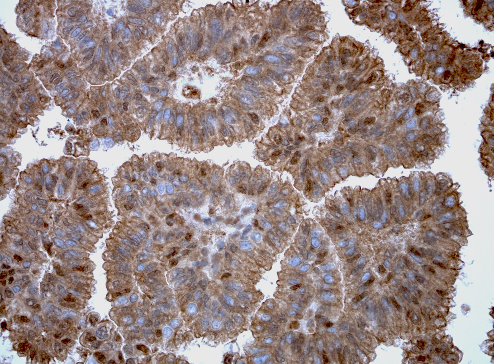
Lane 3: Molt-4 whole cell lysate, 10 µg (input)ARG65859 anti-CD3 epsilon antibody [SQab1713] IHC-P image
Immunohistochemistry: Formalin/PFA-fixed and paraffin-embedded sections of Human colon tissue stained with ARG65859 anti-CD3 epsilon antibody [SQab1713] at 1:200 dilution. Antigen Retrieval: Boil tissue section in Tris/EDTA buffer (pH 9.0).


 New Products