


anti-CST3 / Cystatin C antibody


Key features and details
- 产品描述:
- 反应物种:
- 应用:
- 特异性:
- 宿主:
- 克隆:
- 克隆号:
- 靶点名称:
- 抗原物种:
-
Brand:

 Product Details
| 产品描述 | Mouse Monoclonal antibody recognizes CST3 / Cystatin C |
|---|---|
| 反应物种 | Hu |
| 应用 | IHC-P |
| 特异性 | The antibody detects endogenous Cystatin 3. |
| 宿主 | Mouse |
| 克隆 | Monoclonal |
| 克隆号 | 7F11 |
| 靶点名称 | CST3 / Cystatin C |
| 抗原物种 | Human |
| 抗原 | Recombinant Protein of Human Cystatin 3. |
| 偶联标记 | Un-conjugated |
| 別名 | Cystatin-C; Neuroendocrine basic polypeptide; Post-gamma-globulin; ARMD11; Cystatin-3; Gamma-trace |
| 应用建议 |
| ||||
|---|---|---|---|---|---|
| 应用说明 | IHC-P: Antigen Retrieval: Boil tissue section in Sodium citrate buffer (pH 6.0) for 20 min. * The dilutions indicate recommended starting dilutions and the optimal dilutions or concentrations should be determined by the scientist. |
| 形式 | Liquid |
|---|---|
| 纯化 | Affinity purification with immunogen. |
| 缓冲液 | PBS, 0.02% Sodium azide, 50% Glycerol and 0.5% BSA. |
| 抗菌剂 | 0.02% Sodium azide |
| 稳定剂 | 50% Glycerol and 0.5% BSA |
| 浓度 | 1 mg/ml |
| 存放说明 | For continuous use, store undiluted antibody at 2-8°C for up to a week. For long-term storage, aliquot and store at -20°C. Storage in frost free freezers is not recommended. Avoid repeated freeze/thaw cycles. Suggest spin the vial prior to opening. The antibody solution should be gently mixed before use. |
| 注意事项 | For laboratory research only, not for drug, diagnostic or other use. |
| 数据库连接 | |
|---|---|
| 基因名称 | CST3 |
| 全名 | cystatin C |
| 背景介绍 | The cystatin superfamily encompasses proteins that contain multiple cystatin-like sequences. Some of the members are active cysteine protease inhibitors, while others have lost or perhaps never acquired this inhibitory activity. There are three inhibitory families in the superfamily, including the type 1 cystatins (stefins), type 2 cystatins and the kininogens. The type 2 cystatin proteins are a class of cysteine proteinase inhibitors found in a variety of human fluids and secretions, where they appear to provide protective functions. The cystatin locus on chromosome 20 contains the majority of the type 2 cystatin genes and pseudogenes. This gene is located in the cystatin locus and encodes the most abundant extracellular inhibitor of cysteine proteases, which is found in high concentrations in biological fluids and is expressed in virtually all organs of the body. A mutation in this gene has been associated with amyloid angiopathy. Expression of this protein in vascular wall smooth muscle cells is severely reduced in both atherosclerotic and aneurysmal aortic lesions, establishing its role in vascular disease. In addition, this protein has been shown to have an antimicrobial function, inhibiting the replication of herpes simplex virus. Alternative splicing results in multiple transcript variants encoding a single protein. [provided by RefSeq, Nov 2014] |
| 生物功能 | As an inhibitor of cysteine proteinases, this protein is thought to serve an important physiological role as a local regulator of this enzyme activity. [UniProt] |
| 预测分子量 | 16 kDa |
| 翻译后修饰 | The Thr-25 variant is O-glycosylated with a core 1 or possibly core 8 glycan. The signal peptide of the O-glycosylated Thr-25 variant is cleaved between Ala-20 and Val-21. |
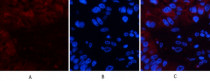
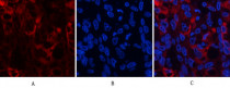
ARG66182 anti-CST3 / Cystatin C antibody IHC image
Immunohistochemistry: Human liver cancer tissue stained with ARG66182 anti-CST3 / Cystatin C antibody (red) at 1:200 dilution (4°C, overnight).
Picture A: Target. Picture B: DAPI. Picture C: merge of A+B.
ARG66182 anti-CST3 / Cystatin C antibody IHC image
Immunohistochemistry: Human liver cancer tissue stained with ARG66182 anti-CST3 / Cystatin C antibody (red) at 1:200 dilution (4°C, overnight).
Picture A: Target. Picture B: DAPI. Picture C: merge of A+B.
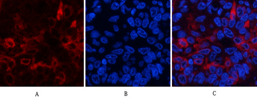
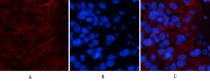
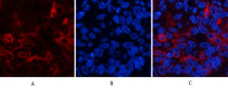
ARG66182 anti-CST3 / Cystatin C antibody IHC image
Immunohistochemistry: Human lung cancer tissue stained with ARG66182 anti-CST3 / Cystatin C antibody (red) at 1:200 dilution (4°C, overnight).
Picture A: Target. Picture B: DAPI. Picture C: merge of A+B.
ARG66182 anti-CST3 / Cystatin C antibody IHC image
Immunohistochemistry: Human lung cancer tissue stained with ARG66182 anti-CST3 / Cystatin C antibody (red) at 1:200 dilution (4°C, overnight).
Picture A: Target. Picture B: DAPI. Picture C: merge of A+B.
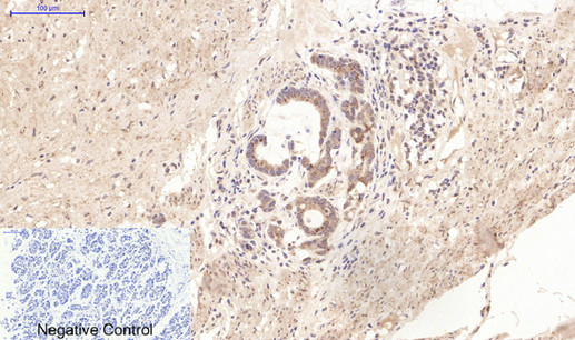
ARG66182 anti-CST3 / Cystatin C antibody IHC-P image
Immunohistochemistry: Paraffin-embedded Human liver tissue stained with ARG66182 anti-CST3 / Cystatin C antibody at 1:200 dilution (4°C, overnight). Antigen Retrieval: Boil tissue section in Sodium citrate buffer (pH 6.0) for 20 min.
Negative control was used by secondary antibody only.
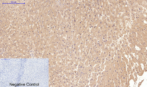
ARG66182 anti-CST3 / Cystatin C antibody IHC-P image
Immunohistochemistry: Paraffin-embedded Human liver cancer tissue stained with ARG66182 anti-CST3 / Cystatin C antibody at 1:200 dilution (4°C, overnight). Antigen Retrieval: Boil tissue section in Sodium citrate buffer (pH 6.0) for 20 min.
Negative control was used by secondary antibody only.

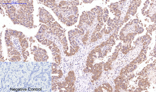
ARG66182 anti-CST3 / Cystatin C antibody IHC-P image
Immunohistochemistry: Paraffin-embedded Human lung cancer tissue stained with ARG66182 anti-CST3 / Cystatin C antibody at 1:200 dilution (4°C, overnight). Antigen Retrieval: Boil tissue section in Sodium citrate buffer (pH 6.0) for 20 min.
Negative control was used by secondary antibody only.
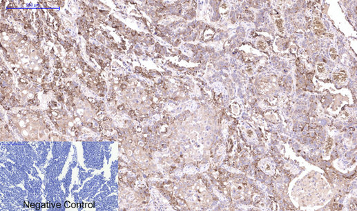
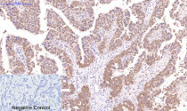
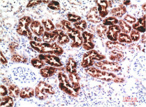
ARG66182 anti-CST3 / Cystatin C antibody IHC-P image
Immunohistochemistry: Paraffin-embedded Human kidney tissue stained with ARG66182 anti-CST3 / Cystatin C antibody at 1:200 dilution (4°C, overnight). Antigen Retrieval: Boil tissue section in Sodium citrate buffer (pH 6.0) for 20 min.
Negative control was used by secondary antibody only.

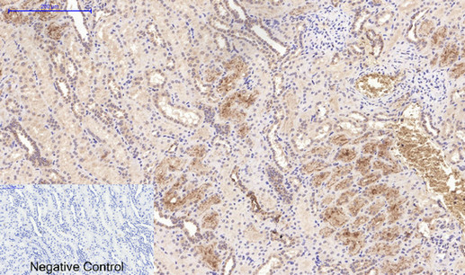
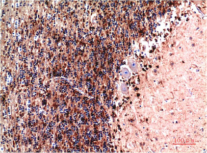
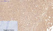
ARG66182 anti-CST3 / Cystatin C antibody IHC-P image
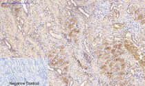
Immunohistochemistry: Paraffin-embedded Human stomach cancer tissue stained with ARG66182 anti-CST3 / Cystatin C antibody at 1:200 dilution (4°C, overnight). Antigen Retrieval: Boil tissue section in Sodium citrate buffer (pH 6.0) for 20 min.
Negative control was used by secondary antibody only.
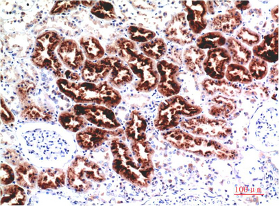
ARG66182 anti-CST3 / Cystatin C antibody IHC-P image
Immunohistochemistry: Paraffin-embedded Human Kidney Tissue stained with ARG66182 anti-CST3 / Cystatin C antibody at 1:200 dilution.
ARG66182 anti-CST3 / Cystatin C antibody IHC-P image
Immunohistochemistry: Paraffin-embedded Human Brain Tissue stained with ARG66182 anti-CST3 / Cystatin C antibody at 1:200 dilution.


 New Products


