


anti-DAG1 antibody

Key features and details
- 产品描述:
- 反应物种:
- 预测物种:
- 应用:
- 特异性:
- 宿主:
- 克隆:
- 同位型:
- 靶点名称:
-
Brand:

 Product Details
| 产品描述 | Goat Polyclonal antibody recognizes DAG1 |
|---|---|
| 反应物种 | Hu, Ms, Rat |
| 预测物种 | Cow, Dog |
| 应用 | IHC-P, WB |
| 特异性 | This antibody is expected to recognize both the precursor and the mature alpha-dystroglycan, but not the mature beta-dystroglycan. |
| 宿主 | Goat |
| 克隆 | Polyclonal |
| 同位型 | IgG |
| 靶点名称 | DAG1 |
| 抗原物种 | Human |
| 抗原 | C-HVGKHEYFMHATDK |
| 偶联标记 | Un-conjugated |
| 別名 | Dystrophin-associated glycoprotein 1; Alpha-DG; DAG; 156DAG; AGRNR; Dystroglycan; A3a; MDDGC9; MDDGC7; Beta-DG |
| 应用建议 |
| ||||||
|---|---|---|---|---|---|---|---|
| 应用说明 | WB: Recommend incubate at RT for 1h. IHC-P: Antigen Retrieval: Steam tissue section in Citrate buffer (pH 6.0). * The dilutions indicate recommended starting dilutions and the optimal dilutions or concentrations should be determined by the scientist. |
| 形式 | Liquid |
|---|---|
| 纯化 | Purified from goat serum by ammonium sulphate precipitation followed by antigen affinity chromatography using the immunizing peptide. |
| 缓冲液 | Tris saline (pH 7.3), 0.02% Sodium azide and 0.5% BSA |
| 抗菌剂 | 0.02% Sodium azide |
| 稳定剂 | 0.5% BSA |
| 浓度 | 0.5 mg/ml |
| 存放说明 | For continuous use, store undiluted antibody at 2-8°C for up to a week. For long-term storage, aliquot and store at -20°C or below. Storage in frost free freezers is not recommended. Avoid repeated freeze/thaw cycles. Suggest spin the vial prior to opening. The antibody solution should be gently mixed before use. |
| 注意事项 | For laboratory research only, not for drug, diagnostic or other use. |
| 数据库连接 | |
|---|---|
| 背景介绍 | Dystroglycan is a laminin binding component of the dystrophin-glycoprotein complex which provides a linkage between the subsarcolemmal cytoskeleton and the extracellular matrix. Dystroglycan 1 is a candidate gene for the site of the mutation in autosomal recessive muscular dystrophies. The dramatic reduction of dystroglycan 1 in Duchenne muscular dystrophy leads to a loss of linkage between the sarcolemma and extracellular matrix, rendering muscle fibers more susceptible to necrosis. Dystroglycan also functions as dual receptor for agrin and laminin-2 in the Schwann cell membrane. The muscle and nonmuscle isoforms of dystroglycan differ by carbohydrate moieties but not protein sequence. Alternative splicing results in multiple transcript variants all encoding the same protein.[provided by RefSeq, Apr 2010] |
| 研究领域 | Neuroscience antibody; Signaling Transduction antibody |
| 预测分子量 | 97 kDa |
| 翻译后修饰 | O- and N-glycosylated. Alpha-dystroglycan is heavily O-glycosylated comprising of up to two thirds of its mass and the carbohydrate composition differs depending on tissue type. Mucin-type O-glycosylation is important for ligand binding activity. O-mannosylation of alpha-DAG1 is found in high abundance in both brain and muscle where the most abundant glycan is Sia-alpha-2-3-Gal-beta-1-4-Glc-NAc-beta-1-2-Man. In muscle, glycosylation on Thr-317, Thr-319 and Thr-379 by a phosphorylated O-mannosyl glycan with the structure 2-(N-acetylamido)-2-deoxygalactosyl-beta-1,3-2-(N-acetylamido)-2-deoxyglucosyl-beta-1,4-6-phosphomannose is mediated by like-acetylglucosaminyltransferase (LARGE1) protein and is required for laminin binding (PubMed:20044576, PubMed:21987822, PubMed:24256719). O-mannosylation is also required for binding lymphocytic choriomeningitis virus, Old World Lassa fever virus, and clade C New World arenaviruses. The O-glycosyl hexose on Thr-367, Thr-369, Thr-372, Thr-381 and Thr-388 is probably mannose. O-glycosylated in the N-terminal region with a core 1 or possibly core 8 glycan. The beta subunit is N-glycosylated. Autolytic cleavage produces the alpha and beta subunits. In cutaneous cells, as well as in certain pathological conditions, shedding of beta-dystroglcan can occur releasing a peptide of about 30 kDa. SRC-mediated phosphorylation of the PPXY motif of the beta subunit recruits SH2 domain-containing proteins, but inhibits binding to WWW domain-containing proteins, DMD and UTRN. This phosphorylation also inhibits nuclear entry. |

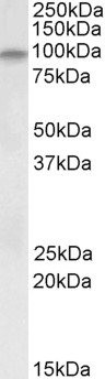
ARG65250 anti-DAG1 antibody WB image
Western Blot: Human Skeletal Muscle lysate (35µg protein in RIPA buffer) stained with ARG65250 anti-DAG1 antibody (0.2µg/ml)

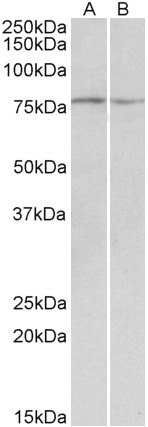
ARG65250 anti-DAG1 antibody WB image
Western Blot: Mouse and Rat Skeletal Muscle lysate (35µg protein in RIPA buffer) stained with ARG65250 anti-DAG1 antibody (0.1µg/ml)
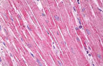
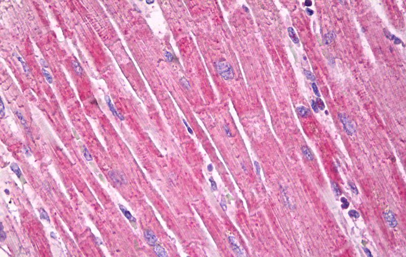
ARG65250 anti-DAG1 antibody IHC-P image
Immunohistochemistry: Paraffin-embedded Human heart tissue. Antigen Retrieval: Steam tissue section in Citrate buffer (pH 6.0). The tissue section was stained with ARG65250 anti-DAG1 antibody at 5 µg/ml dilution followed by AP-staining.


High-Intensity Interval Training Attenuates Ketogenic Diet-Induced Liver Fibrosis in Type 2 Diabetic Mice by Ameliorating TGF-β1/Smad Signaling.
WB / Mouse

 New Products


