


anti-DBP / Vitamin D binding protein antibody


Key features and details
- 产品描述:
- 反应物种:
- 预测物种:
- 应用:
- 宿主:
- 克隆:
- 同位型:
- 靶点名称:
- 抗原物种:
-
Brand:

 Product Details
| 产品描述 | Rabbit Polyclonal antibody recognizes DBP / Vitamin D binding protein |
|---|---|
| 反应物种 | Hu, Ms |
| 预测物种 | Rat |
| 应用 | IHC-P, WB |
| 宿主 | Rabbit |
| 克隆 | Polyclonal |
| 同位型 | IgG |
| 靶点名称 | DBP / Vitamin D binding protein |
| 抗原物种 | Human |
| 抗原 | Recombinant protein corresponding to L17-E256 of Human DBP. |
| 偶联标记 | Un-conjugated |
| 別名 | GRD3; DBP/GC; HEL-S-51; VDBG; VDB; Gc-globulin; DBP; VDBP; Vitamin D-binding protein; Group-specific component |
| 应用建议 |
| ||||||
|---|---|---|---|---|---|---|---|
| 应用说明 | IHC-P: Antigen Retrieval: Heat mediation was performed in Citrate buffer (pH 6.0, epitope retrieval solution) for 20 min. * The dilutions indicate recommended starting dilutions and the optimal dilutions or concentrations should be determined by the scientist. |
| 形式 | Liquid |
|---|---|
| 纯化 | Affinity purification with immunogen. |
| 缓冲液 | 0.2% Na2HPO4, 0.9% NaCl, 0.05% Sodium azide and 5% BSA. |
| 抗菌剂 | 0.05% Sodium azide |
| 稳定剂 | 5% BSA |
| 浓度 | 0.5 mg/ml |
| 存放说明 | For continuous use, store undiluted antibody at 2-8°C for up to a week. For long-term storage, aliquot and store at -20°C or below. Storage in frost free freezers is not recommended. Avoid repeated freeze/thaw cycles. Suggest spin the vial prior to opening. The antibody solution should be gently mixed before use. |
| 注意事项 | For laboratory research only, not for drug, diagnostic or other use. |
| 数据库连接 | |
|---|---|
| 基因名称 | GC |
| 全名 | group-specific component (vitamin D binding protein) |
| 背景介绍 | The protein encoded by this gene belongs to the albumin gene family. It is a multifunctional protein found in plasma, ascitic fluid, cerebrospinal fluid and on the surface of many cell types. It binds to vitamin D and its plasma metabolites and transports them to target tissues. Alternatively spliced transcript variants encoding different isoforms have been found for this gene.[provided by RefSeq, Feb 2011] |
| 生物功能 | Multifunctional protein found in plasma, ascitic fluid, cerebrospinal fluid, and urine and on the surface of many cell types. In plasma, it carries the vitamin D sterols and prevents polymerization of actin by binding its monomers. DBP associates with membrane-bound immunoglobulin on the surface of B-lymphocytes and with IgG Fc receptor on the membranes of T-lymphocytes. [UniProt] |
| 细胞定位 | Secreted. [UniProt] |
| 预测分子量 | 53 kDa |
| 翻译后修饰 | Allele GC*1S is O-glycosylated at Thr-436 (PubMed:20079467). The trisaccharide sugar moiety can be modified by the successive removal of neuraminic acid and galactose leaving an O-linked N-acetyl-galactosamine. This conversion is thought to produce a macrophage-activating factor (Gc-MAF). Only a minor proportion of plasma GC is O-glycosylated (PubMed:17360250). The potential N-glycosylation site predicted at Asn-288 is thought to be nonglycosylated. [UniProt] |
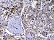
ARG40921 anti-DBP / Vitamin D binding protein antibody IHC-P image
Immunohistochemistry: Paraffin-embedded Human liver cancer tissue. Antigen Retrieval: Heat mediation was performed in Citrate buffer (pH 6.0, epitope retrieval solution) for 20 min. The tissue section was blocked with 10% goat serum. The tissue section was then stained with ARG40921 anti-DBP / Vitamin D binding protein antibody at 1 µg/ml dilution, overnight at 4°C.

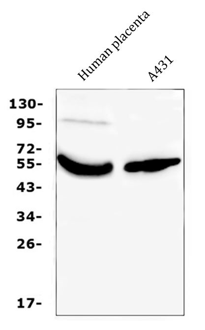
ARG40921 anti-DBP / Vitamin D binding protein antibody WB image
Western blot: 50 µg of samples under reducing conditions. Human placenta and A431 whole cell lysates stained with ARG40921 anti-DBP / Vitamin D binding protein antibody at 0.5 µg/ml, overnight at 4°C.
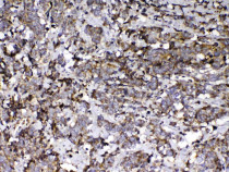
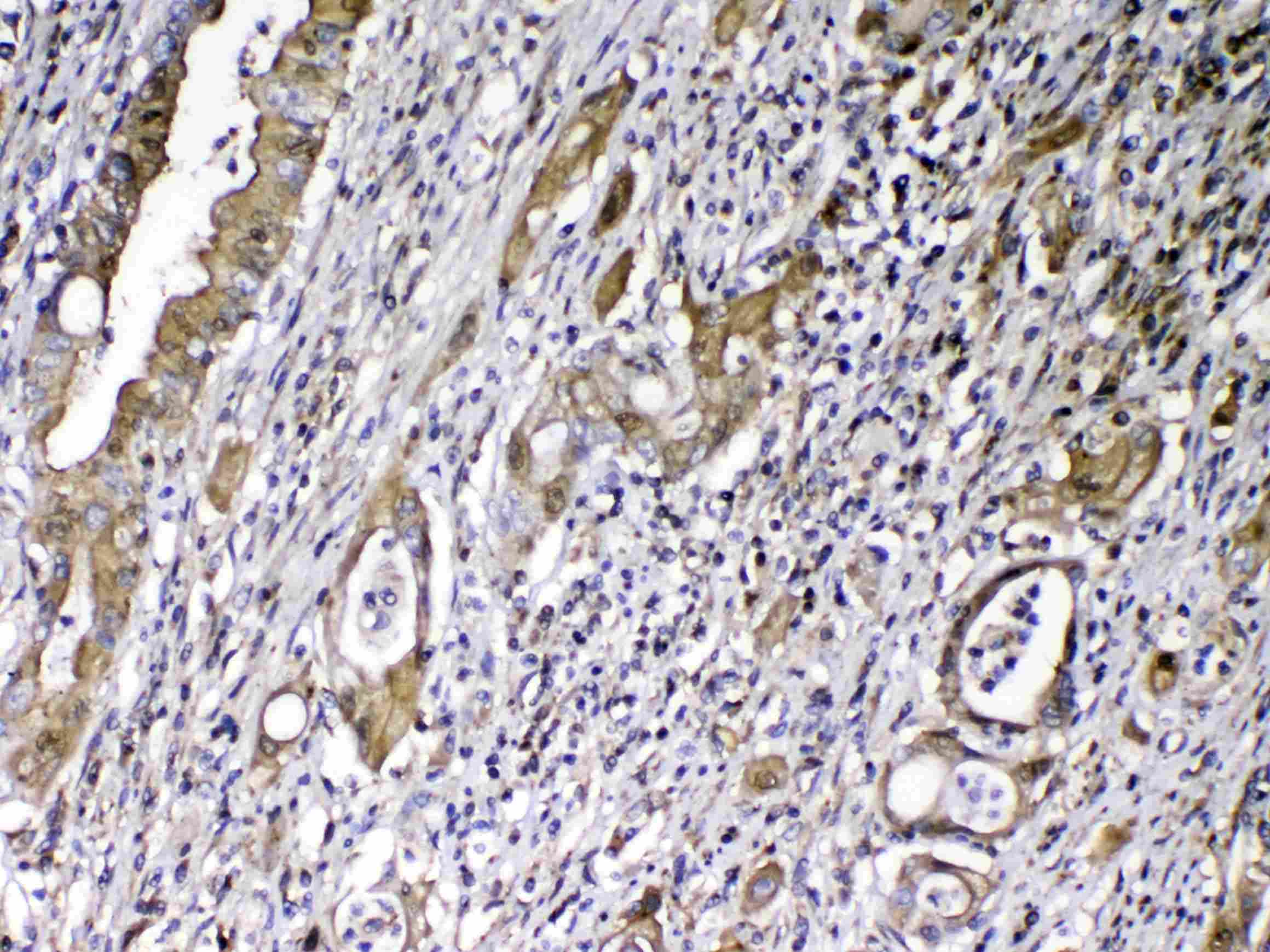
ARG40921 anti-DBP / Vitamin D binding protein antibody IHC-P image
Immunohistochemistry: Paraffin-embedded Human lung cancer tissue. Antigen Retrieval: Heat mediation was performed in Citrate buffer (pH 6.0, epitope retrieval solution) for 20 min. The tissue section was blocked with 10% goat serum. The tissue section was then stained with ARG40921 anti-DBP / Vitamin D binding protein antibody at 1 µg/ml dilution, overnight at 4°C.
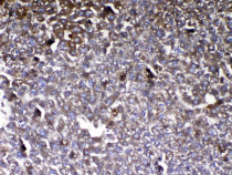
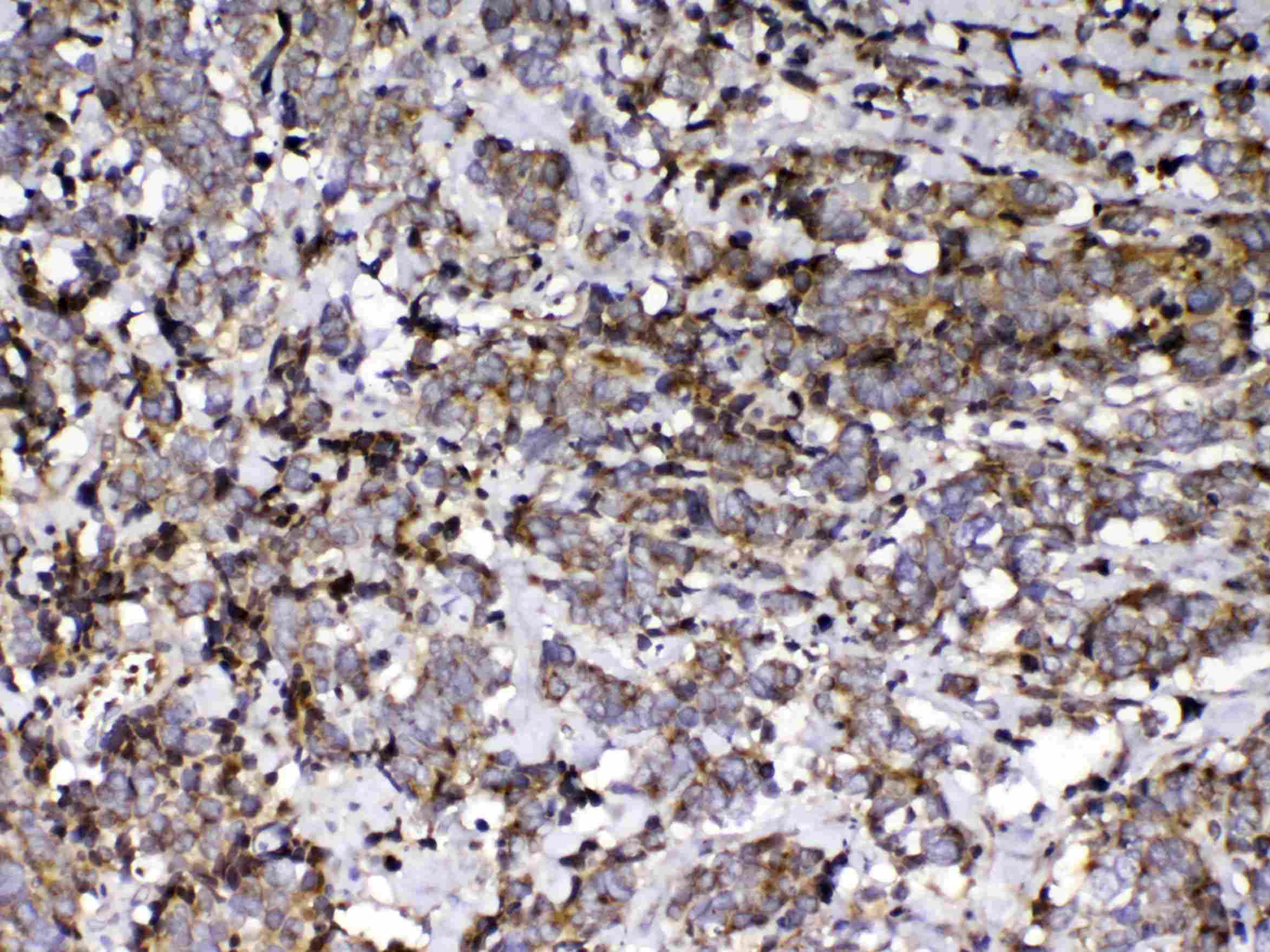
ARG40921 anti-DBP / Vitamin D binding protein antibody IHC-P image
Immunohistochemistry: Paraffin-embedded Mouse liver tissue. Antigen Retrieval: Heat mediation was performed in Citrate buffer (pH 6.0, epitope retrieval solution) for 20 min. The tissue section was blocked with 10% goat serum. The tissue section was then stained with ARG40921 anti-DBP / Vitamin D binding protein antibody at 1 µg/ml dilution, overnight at 4°C.
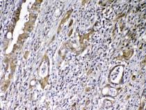
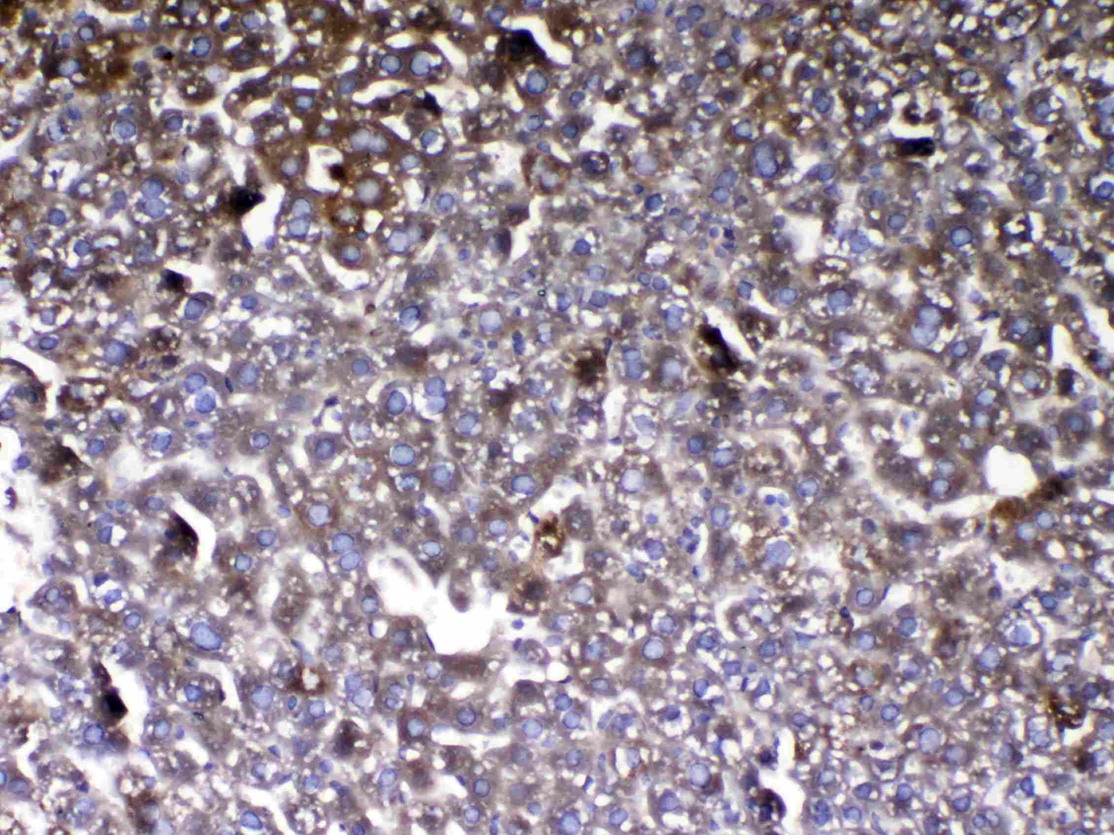
ARG40921 anti-DBP / Vitamin D binding protein antibody IHC-P image
Immunohistochemistry: Paraffin-embedded Human rectal cancer tissue. Antigen Retrieval: Heat mediation was performed in Citrate buffer (pH 6.0, epitope retrieval solution) for 20 min. The tissue section was blocked with 10% goat serum. The tissue section was then stained with ARG40921 anti-DBP / Vitamin D binding protein antibody at 1 µg/ml dilution, overnight at 4°C.


 New Products


