


anti-EFHD1 antibody


Key features and details
- 产品描述:
- 反应物种:
- 应用:
- 特异性:
- 宿主:
- 克隆:
- 克隆号:
- 靶点名称:
- 抗原物种:
-
Brand:

 Product Details
| 产品描述 | Mouse Monoclonal antibody recognizes EFHD1 |
|---|---|
| 反应物种 | Hu, Ms, Rat |
| 应用 | ICC/IF, IHC-P |
| 特异性 | The antibody detects endogenous EFHD1 proteins. |
| 宿主 | Mouse |
| 克隆 | Monoclonal |
| 克隆号 | 3G2 |
| 靶点名称 | EFHD1 |
| 抗原物种 | Human |
| 抗原 | Synthetic peptide of Human EFHD1. |
| 偶联标记 | Un-conjugated |
| 別名 | EF-hand domain-containing protein D1; Swiprosin-2; EF-hand domain-containing protein 1; MSTP133; MST133; SWS2; PP3051 |
| 应用建议 |
| ||||||
|---|---|---|---|---|---|---|---|
| 应用说明 | IHC-P: Antigen Retrieval: Boil tissue section in Sodium citrate buffer (pH 6.0) for 20 min. * The dilutions indicate recommended starting dilutions and the optimal dilutions or concentrations should be determined by the scientist. |
| 形式 | Liquid |
|---|---|
| 纯化 | Affinity purification with immunogen. |
| 缓冲液 | PBS (pH 7.4), 0.02% Sodium azide and 50% Glycerol. |
| 抗菌剂 | 0.02% Sodium azide |
| 稳定剂 | 50% Glycerol |
| 浓度 | 1 mg/ml |
| 存放说明 | For continuous use, store undiluted antibody at 2-8°C for up to a week. For long-term storage, aliquot and store at -20°C. Storage in frost free freezers is not recommended. Avoid repeated freeze/thaw cycles. Suggest spin the vial prior to opening. The antibody solution should be gently mixed before use. |
| 注意事项 | For laboratory research only, not for drug, diagnostic or other use. |
| 数据库连接 | Swiss-port # Q9BUP0 Human EF-hand domain-containing protein D1 Swiss-port # Q9D4J1 Mouse EF-hand domain-containing protein D1 |
|---|---|
| 基因名称 | EFHD1 |
| 全名 | EF-hand domain family, member D1 |
| 背景介绍 | EFHD1 is an EF-hand domain-containing protein that displays increased expression during neuronal differentiation (Tominaga and Tomooka, 2002 [PubMed 12270117]).[supplied by OMIM, Mar 2008] |
| 预测分子量 | 27 kDa |
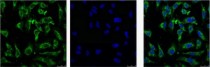
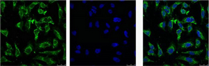
ARG66174 anti-EFHD1 antibody ICC/IF image
Immunofluorescence: HeLa cells stained with ARG66174 anti-EFHD1 antibody (green).
Left: Target. Middle: DAPI. Right: merge image.
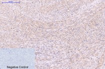
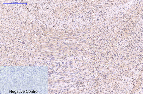
ARG66174 anti-EFHD1 antibody IHC-P image
Immunohistochemistry: Paraffin-embedded Human uterus tissue stained with ARG66174 anti-EFHD1 antibody at 1:200 dilution (4°C, overnight). Antigen Retrieval: Boil tissue section in Sodium citrate buffer (pH 6.0) for 20 min.
Negative control was used by secondary antibody only.
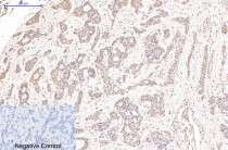
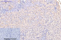
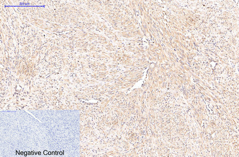
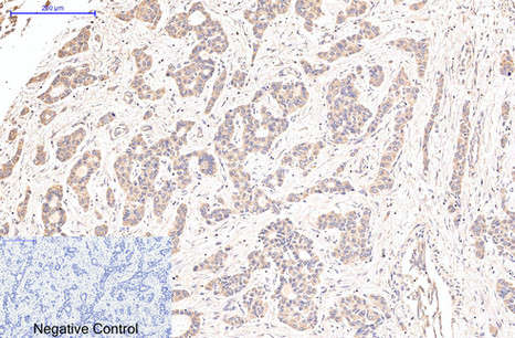
ARG66174 anti-EFHD1 antibody IHC-P image
Immunohistochemistry: Paraffin-embedded Human uterus cancer tissue stained with ARG66174 anti-EFHD1 antibody at 1:200 dilution (4°C, overnight). Antigen Retrieval: Boil tissue section in Sodium citrate buffer (pH 6.0) for 20 min.
Negative control was used by secondary antibody only.
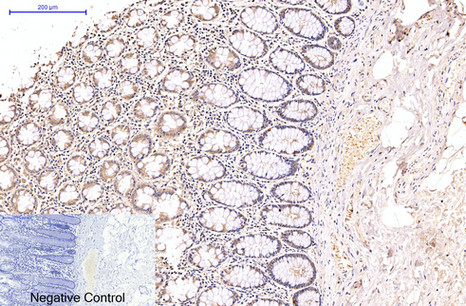
ARG66174 anti-EFHD1 antibody IHC-P image
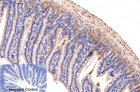
Immunohistochemistry: Paraffin-embedded Human colon tissue stained with ARG66174 anti-EFHD1 antibody at 1:200 dilution (4°C, overnight). Antigen Retrieval: Boil tissue section in Sodium citrate buffer (pH 6.0) for 20 min.
Negative control was used by secondary antibody only.

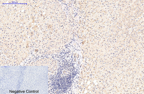
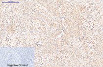
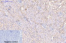
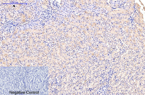
ARG66174 anti-EFHD1 antibody IHC-P image
Immunohistochemistry: Paraffin-embedded Human liver tissue stained with ARG66174 anti-EFHD1 antibody at 1:200 dilution (4°C, overnight). Antigen Retrieval: Boil tissue section in Sodium citrate buffer (pH 6.0) for 20 min.
Negative control was used by secondary antibody only.
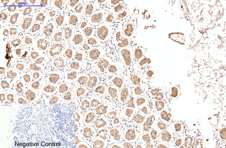
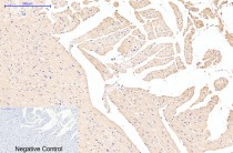
ARG66174 anti-EFHD1 antibody IHC-P image
Immunohistochemistry: Paraffin-embedded Human liver cancer tissue stained with ARG66174 anti-EFHD1 antibody at 1:200 dilution (4°C, overnight). Antigen Retrieval: Boil tissue section in Sodium citrate buffer (pH 6.0) for 20 min.
Negative control was used by secondary antibody only.

ARG66174 anti-EFHD1 antibody IHC-P image
Immunohistochemistry: Paraffin-embedded Human stomach tissue stained with ARG66174 anti-EFHD1 antibody at 1:200 dilution (4°C, overnight). Antigen Retrieval: Boil tissue section in Sodium citrate buffer (pH 6.0) for 20 min.
Negative control was used by secondary antibody only.
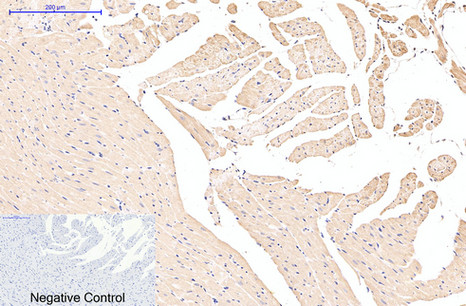
ARG66174 anti-EFHD1 antibody IHC-P image
Immunohistochemistry: Paraffin-embedded Rat heart tissue stained with ARG66174 anti-EFHD1 antibody at 1:200 dilution (4°C, overnight). Antigen Retrieval: Boil tissue section in Sodium citrate buffer (pH 6.0) for 20 min.
Negative control was used by secondary antibody only.

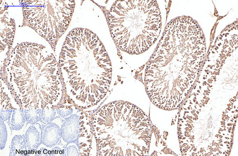
ARG66174 anti-EFHD1 antibody IHC-P image
Immunohistochemistry: Paraffin-embedded Rat testis tissue stained with ARG66174 anti-EFHD1 antibody at 1:200 dilution (4°C, overnight). Antigen Retrieval: Boil tissue section in Sodium citrate buffer (pH 6.0) for 20 min.
Negative control was used by secondary antibody only.
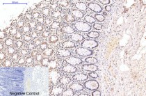
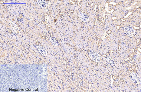
ARG66174 anti-EFHD1 antibody IHC-P image
Immunohistochemistry: Paraffin-embedded Rat kidney tissue stained with ARG66174 anti-EFHD1 antibody at 1:200 dilution (4°C, overnight). Antigen Retrieval: Boil tissue section in Sodium citrate buffer (pH 6.0) for 20 min.
Negative control was used by secondary antibody only.
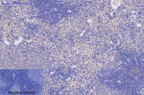
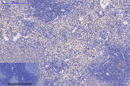
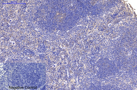
ARG66174 anti-EFHD1 antibody IHC-P image
Immunohistochemistry: Paraffin-embedded Rat spleen tissue stained with ARG66174 anti-EFHD1 antibody at 1:200 dilution (4°C, overnight). Antigen Retrieval: Boil tissue section in Sodium citrate buffer (pH 6.0) for 20 min.
Negative control was used by secondary antibody only.
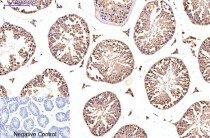
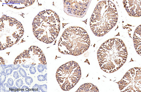
ARG66174 anti-EFHD1 antibody IHC-P image
Immunohistochemistry: Paraffin-embedded Mouse testis tissue stained with ARG66174 anti-EFHD1 antibody at 1:200 dilution (4°C, overnight). Antigen Retrieval: Boil tissue section in Sodium citrate buffer (pH 6.0) for 20 min.
Negative control was used by secondary antibody only.

ARG66174 anti-EFHD1 antibody IHC-P image
Immunohistochemistry: Paraffin-embedded Mouse colon tissue stained with ARG66174 anti-EFHD1 antibody at 1:200 dilution (4°C, overnight). Antigen Retrieval: Boil tissue section in Sodium citrate buffer (pH 6.0) for 20 min.
Negative control was used by secondary antibody only.
ARG66174 anti-EFHD1 antibody IHC-P image
Immunohistochemistry: Paraffin-embedded Mouse kidney tissue stained with ARG66174 anti-EFHD1 antibody at 1:200 dilution (4°C, overnight). Antigen Retrieval: Boil tissue section in Sodium citrate buffer (pH 6.0) for 20 min.
Negative control was used by secondary antibody only.
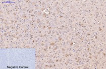
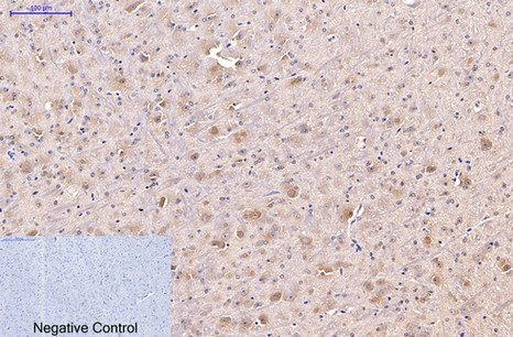
ARG66174 anti-EFHD1 antibody IHC-P image
Immunohistochemistry: Paraffin-embedded Mouse brain tissue stained with ARG66174 anti-EFHD1 antibody at 1:200 dilution (4°C, overnight). Antigen Retrieval: Boil tissue section in Sodium citrate buffer (pH 6.0) for 20 min.
Negative control was used by secondary antibody only.

ARG66174 anti-EFHD1 antibody IHC-P image
Immunohistochemistry: Paraffin-embedded Mouse spleen tissue stained with ARG66174 anti-EFHD1 antibody at 1:200 dilution (4°C, overnight). Antigen Retrieval: Boil tissue section in Sodium citrate buffer (pH 6.0) for 20 min.
Negative control was used by secondary antibody only.
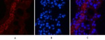
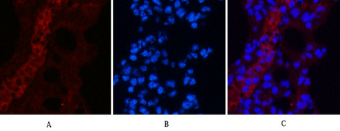
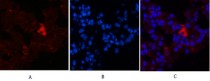
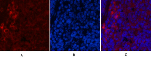
ARG66174 anti-EFHD1 antibody IHC image
Immunohistochemistry: Mouse lung tissue stained with ARG66174 anti-EFHD1 antibody (red) at 1:200 dilution (4°C, overnight).
Picture A: Target. Picture B: DAPI. Picture C: merge of A+B.
ARG66174 anti-EFHD1 antibody IHC image
Immunohistochemistry: Mouse lung tissue stained with ARG66174 anti-EFHD1 antibody (red) at 1:200 dilution (4°C, overnight).
Picture A: Target. Picture B: DAPI. Picture C: merge of A+B.
ARG66174 anti-EFHD1 antibody IHC image
Immunohistochemistry: Mouse lung tissue stained with ARG66174 anti-EFHD1 antibody (red) at 1:200 dilution (4°C, overnight).
Picture A: Target. Picture B: DAPI. Picture C: merge of A+B.
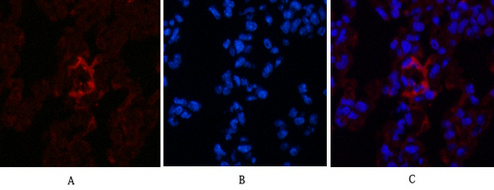
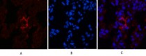
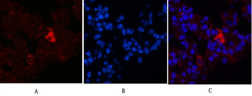
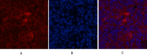
ARG66174 anti-EFHD1 antibody IHC image
Immunohistochemistry: Mouse spleen tissue stained with ARG66174 anti-EFHD1 antibody (red) at 1:200 dilution (4°C, overnight).
Picture A: Target. Picture B: DAPI. Picture C: merge of A+B.
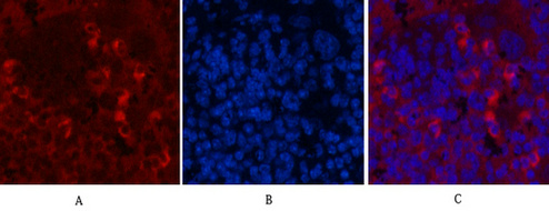
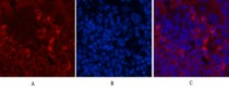
ARG66174 anti-EFHD1 antibody IHC image
Immunohistochemistry: Mouse spleen tissue stained with ARG66174 anti-EFHD1 antibody (red) at 1:200 dilution (4°C, overnight).
Picture A: Target. Picture B: DAPI. Picture C: merge of A+B.
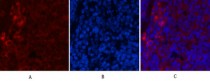
ARG66174 anti-EFHD1 antibody IHC image
Immunohistochemistry: Mouse spleen tissue stained with ARG66174 anti-EFHD1 antibody (red) at 1:200 dilution (4°C, overnight).
Picture A: Target. Picture B: DAPI. Picture C: merge of A+B.


 New Products


