


anti-EHD1 antibody


Key features and details
- 产品描述:
- 反应物种:
- 预测物种:
- 应用:
- 宿主:
- 克隆:
- 同位型:
- 靶点名称:
- 抗原物种:
-
Brand:

 Product Details
| 产品描述 | Goat Polyclonal antibody recognizes EHD1 |
|---|---|
| 反应物种 | Hu, Ms |
| 预测物种 | Cow, Rat |
| 应用 | FACS, ICC/IF, IHC-P, WB |
| 宿主 | Goat |
| 克隆 | Polyclonal |
| 同位型 | IgG |
| 靶点名称 | EHD1 |
| 抗原物种 | Human |
| 抗原 | Synthetic peptide around the N-terminus of Human EHD1. (FSWVSKDARRKKEPC) (NP_001269373.1; NP_001269374.1) |
| 偶联标记 | Un-conjugated |
| 別名 | EH domain-containing protein 1; HPAST1; PAST1; Testilin; PAST; hPAST1; H-PAST; PAST homolog 1 |
| 应用建议 |
| ||||||||||
|---|---|---|---|---|---|---|---|---|---|---|---|
| 应用说明 | WB: Recommend incubate at RT for 1h. IHC-P: Antigen Retrieval: Steam tissue section in Citrate buffer (pH 6.0). * The dilutions indicate recommended starting dilutions and the optimal dilutions or concentrations should be determined by the scientist. | ||||||||||
| 阳性对照 | NIH/3T3 | ||||||||||
| 实际分子量 | ~ 63 kDa |
| 形式 | Liquid |
|---|---|
| 纯化 | Ammonium sulphate precipitation followed by affinity purification with immunogen. |
| 缓冲液 | Tris saline (pH 7.3), 0.02% Sodium azide and 0.5% BSA. |
| 抗菌剂 | 0.02% Sodium azide |
| 稳定剂 | 0.5% BSA |
| 浓度 | 0.5 mg/ml |
| 存放说明 | For continuous use, store undiluted antibody at 2-8°C for up to a week. For long-term storage, aliquot and store at -20°C or below. Storage in frost free freezers is not recommended. Avoid repeated freeze/thaw cycles. Suggest spin the vial prior to opening. The antibody solution should be gently mixed before use. |
| 注意事项 | For laboratory research only, not for drug, diagnostic or other use. |
| 数据库连接 | |
|---|---|
| 基因名称 | EHD1 |
| 全名 | EH-domain containing 1 |
| 背景介绍 | This gene belongs to a highly conserved gene family encoding EPS15 homology (EH) domain-containing proteins. The protein-binding EH domain was first noted in EPS15, a substrate for the epidermal growth factor receptor. The EH domain has been shown to be an important motif in proteins involved in protein-protein interactions and in intracellular sorting. The protein encoded by this gene is thought to play a role in the endocytosis of IGF1 receptors. Alternatively spliced transcript variants have been found for this gene. [provided by RefSeq, Sep 2013] |
| 生物功能 | ATP- and membrane-binding protein that controls membrane reorganization/tubulation upon ATP hydrolysis. In vitro causes vesiculation of endocytic membranes (PubMed:24019528). Acts in early endocytic membrane fusion and membrane trafficking of recycling endosomes (PubMed:15020713, PubMed:17233914, PubMed:20801876). Recruited to endosomal membranes upon nerve growth factor stimulation, indirectly regulates neurite outgrowth (By similarity). Plays a role in myoblast fusion (By similarity). Involved in the unidirectional retrograde dendritic transport of endocytosed BACE1 and in efficient sorting of BACE1 to axons implicating a function in neuronal APP processing (By similarity). Plays a role in the formation of the ciliary vesicle (CV), an early step in cilium biogenesis. Proposed to be required for the fusion of distal appendage vesicles (DAVs) to form the CV by recruiting SNARE complex component SNAP29. Is required for recruitment of transition zone proteins CEP290, RPGRIP1L, TMEM67 and B9D2, and of IFT20 following DAV reorganization before Rab8-dependent ciliary membrane extension. Required for the loss of CCP110 form the mother centriole essential for the maturation of the basal body during ciliogenesis (PubMed:25686250). [UniProt] |
| 细胞定位 | Recycling endosome membrane; Peripheral membrane protein; Cytoplasmic side. Early endosome membrane; Peripheral membrane protein; Cytoplasmic side. Cell membrane; Peripheral membrane protein; Cytoplasmic side. Cell projection, cilium membrane; Peripheral membrane protein; Cytoplasmic side. [UniProt] |
| 预测分子量 | 61 kDa |
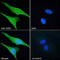
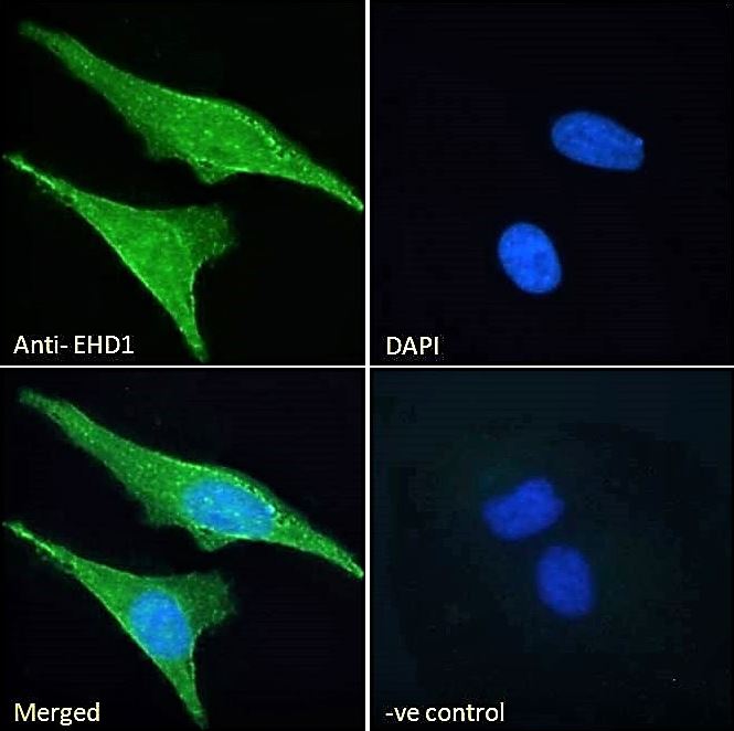
ARG42140 anti-EHD1 antibody ICC/IF image
Immunofluorescence: Paraformaldehyde fixed HeLa cells permeabilized with 0.15% Triton. Cells were stained with ARG42140 anti-EHD1 antibody (green) at 10 µg/ml dilution for 1 hour. DAPI (blue) for nuclear staining. Negative control: Unimmunized Goat IgG at 10 µg/ml dilution.

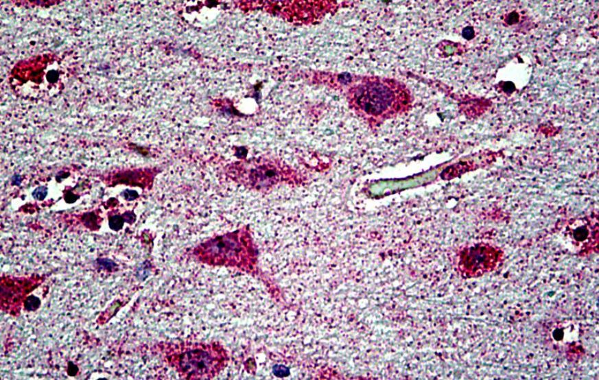
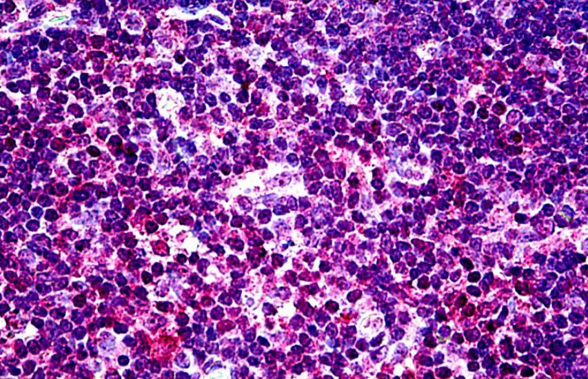
ARG42140 anti-EHD1 antibody IHC-P image
Immunohistochemistry: Paraffin-embedded Human thymus tissue. Antigen Retrieval: Steam tissue section in Citrate buffer (pH 6.0). The tissue section was stained with ARG42140 anti-EHD1 antibody at 3.75 µg/ml dilution followed by AP-staining.
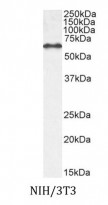
ARG42140 anti-EHD1 antibody WB image
Western blot: 35 µg of NIH/3T3 cell lysate (in RIPA buffer) stained with ARG42140 anti-EHD1 antibody at 0.1 µg/ml dilution and incubated at RT for 1 hour.
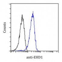
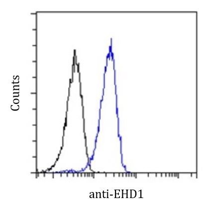
ARG42140 anti-EHD1 antibody FACS image
Flow Cytometry: Paraformaldehyde fixed HeLa cells permeabilized with 0.5% Triton. Cells were stained with ARG42140 anti-EHD1 antibody (blue line) at 10 µg/ml dilution for 1 hour, followed by incubation with Alexa Fluor® 488 labelled secondary antibody. IgG control: Unimmunized Goat IgG (black line) followed by Alexa Fluor® 488 secondary antibody.
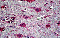
ARG42140 anti-EHD1 antibody IHC-P image
Immunohistochemistry: Paraffin-embedded Human cortex tissue. Antigen Retrieval: Steam tissue section in Citrate buffer (pH 6.0). The tissue section was stained with ARG42140 anti-EHD1 antibody at 3.75 µg/ml dilution followed by AP-staining.

 New Products


