


anti-GFAP antibody


Key features and details
- 产品描述:
- 反应物种:
- 应用:
- 宿主:
- 克隆:
- 同位型:
- 靶点名称:
- 抗原物种:
- 抗原:
-
Brand:

 Product Details
| 产品描述 | Goat Polyclonal antibody recognizes GFAP |
|---|---|
| 反应物种 | Hu, Ms, Rat, Cow |
| 应用 | IHC-Fr, WB |
| 宿主 | Goat |
| 克隆 | Polyclonal |
| 同位型 | IgG |
| 靶点名称 | GFAP |
| 抗原物种 | Human |
| 抗原 | Recombinant full-length Human GFAP isotype 1. |
| 偶联标记 | Un-conjugated |
| 別名 | Glial fibrillary acidic protein; ALXDRD; GFAP |
| 应用建议 |
| ||||||
|---|---|---|---|---|---|---|---|
| 应用说明 | * The dilutions indicate recommended starting dilutions and the optimal dilutions or concentrations should be determined by the scientist. |
| 形式 | Liquid |
|---|---|
| 纯化 | Affinity purified. |
| 缓冲液 | PBS, 5 mM Sodium azide and 50% Glycerol. |
| 抗菌剂 | 5 mM Sodium azide |
| 稳定剂 | 50% Glycerol |
| 浓度 | 1 mg/ml |
| 存放说明 | For continuous use, store undiluted antibody at 2-8°C for up to a week. For long-term storage, aliquot and store at -20°C. Storage in frost free freezers is not recommended. Avoid repeated freeze/thaw cycles. Suggest spin the vial prior to opening. The antibody solution should be gently mixed before use. |
| 注意事项 | For laboratory research only, not for drug, diagnostic or other use. |
| 数据库连接 | |
|---|---|
| 基因名称 | GFAP |
| 全名 | glial fibrillary acidic protein |
| 背景介绍 | This gene encodes one of the major intermediate filament proteins of mature astrocytes. It is used as a marker to distinguish astrocytes from other glial cells during development. Mutations in this gene cause Alexander disease, a rare disorder of astrocytes in the central nervous system. Alternative splicing results in multiple transcript variants encoding distinct isoforms. [provided by RefSeq, Oct 2008] |
| 生物功能 | GFAP, a class-III intermediate filament, is a cell-specific marker that, during the development of the central nervous system, distinguishes astrocytes from other glial cells. [UniProt] |
| 细胞定位 | Cytoplasm. Note=Associated with intermediate filaments. [UniProt] |
| 预测分子量 | 50 kDa |
| 翻译后修饰 | Phosphorylated by PKN1. [UniProt] |

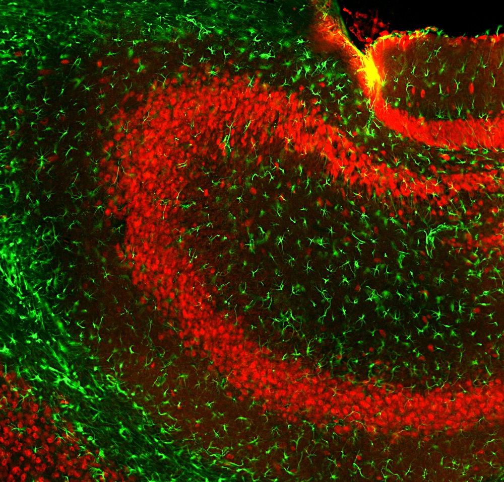
ARG11118 anti-GFAP antibody IHC-Fr image
Immunohistochemistry: Frozen section of Mouse hippocampus tissue stained with ARG11118 anti-GFAP antibody (green) at 1:5000 dilution, and co-stained with ARG52283 anti-FOX3 / NeuN antibody [1B7] (red) at 1:2000 dilution. Hoechst (blue) for nuclear staining. (Sample preparation: Following transcardial perfusion of mouse with 4% paraformaldehyde, brain was post fixed for 24 hours, cut to 45 µM, and free-floating sections were stained with above antibodies.).
ARG11118 anti-GFAP antibody WB image
Western blot: Rat cortex, Rat cerebellum, Mouse cortex, Mouse cerebellum, Cow cortex and Cow cerebellum lysates stained with ARG11118 anti-GFAP antibody at 1:5000 dilution.
Strong band at about 50 kDa corresponds to GFAP protein. Smaller proteolytic fragments of GFAP are also detected on the blot.

 New Products


