


anti-HGD antibody


Key features and details
- 产品描述:
- 反应物种:
- 应用:
- 宿主:
- 克隆:
- 同位型:
- 靶点名称:
- 抗原物种:
- 抗原:
-
Brand:

 Product Details
| 产品描述 | Rabbit Polyclonal antibody recognizes HGD |
|---|---|
| 反应物种 | Hu, Ms, Rat |
| 应用 | FACS, ICC/IF, IHC-P, WB |
| 宿主 | Rabbit |
| 克隆 | Polyclonal |
| 同位型 | IgG |
| 靶点名称 | HGD |
| 抗原物种 | Human |
| 抗原 | Recombinant protein corresponding to D374-N445 of Human HGD. |
| 偶联标记 | Un-conjugated |
| 別名 | EC 1.13.11.5; AKU; HGO; Homogentisate 1,2-dioxygenase; Homogentisate oxygenase; Homogentisicase; Homogentisic acid oxidase |
| 应用建议 |
| ||||||||||
|---|---|---|---|---|---|---|---|---|---|---|---|
| 应用说明 | IHC-P: Antigen Retrieval: Heat mediation was performed in Citrate buffer (pH 6.0) for 20 min. * The dilutions indicate recommended starting dilutions and the optimal dilutions or concentrations should be determined by the scientist. |
| 形式 | Liquid |
|---|---|
| 纯化 | Affinity purification with immunogen. |
| 缓冲液 | 0.9% NaCl, 0.2% Na2HPO4, 0.05% Sodium azide and 4% Trehalose. |
| 抗菌剂 | 0.05% Sodium azide |
| 稳定剂 | 4% Trehalose |
| 浓度 | 0.5 mg/ml |
| 存放说明 | For continuous use, store undiluted antibody at 2-8°C for up to a week. For long-term storage, aliquot and store at -20°C or below. Storage in frost free freezers is not recommended. Avoid repeated freeze/thaw cycles. Suggest spin the vial prior to opening. The antibody solution should be gently mixed before use. |
| 注意事项 | For laboratory research only, not for drug, diagnostic or other use. |
| 数据库连接 | |
|---|---|
| 基因名称 | HGD |
| 全名 | homogentisate 1,2-dioxygenase |
| 背景介绍 | This gene encodes the enzyme homogentisate 1,2 dioxygenase. This enzyme is involved in the catabolism of the amino acids tyrosine and phenylalanine. Mutations in this gene are the cause of the autosomal recessive metabolism disorder alkaptonuria.[provided by RefSeq, May 2010] |
| 预测分子量 | 50 kDa |
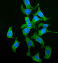
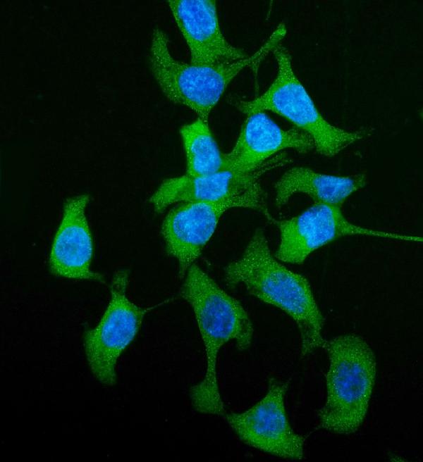
ARG58791 anti-HGD antibody ICC/IF image
Immunofluorescence: Caco-2 cells were blocked with 10% goat serum and then stained with ARG58791 anti-HGD antibody (green) at 5 µg/ml dilution, overnight at 4°C. DAPI (blue) for nuclear staining.

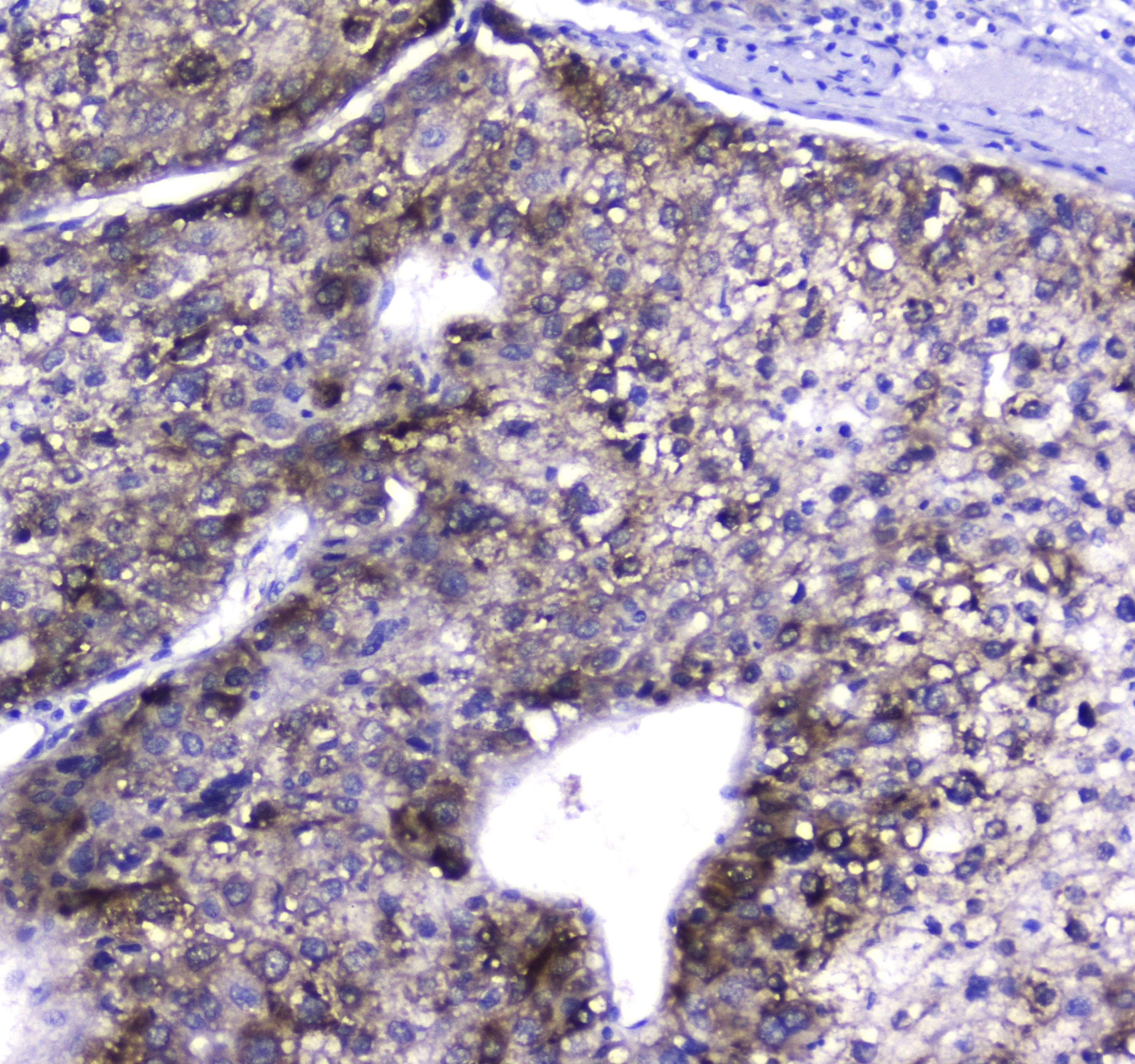
ARG58791 anti-HGD antibody IHC-P image
Immunohistochemistry: Paraffin-embedded Human liver cancer tissue. Antigen Retrieval: Heat mediated was performed in Citrate buffer (pH 6.0, epitope retrieval solution) for 20 min. The tissue section was blocked with 10% goat serum. The tissue section was then stained with ARG58791 anti-HGD antibody at 2 µg/ml, overnight at 4°C.

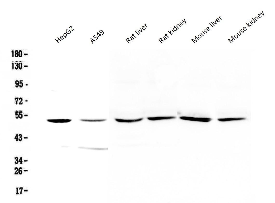
ARG58791 anti-HGD antibody WB image
Western blot: 50 µg of samples under reducing conditions. HepG2, A549, Rat liver, Rat kidney, Mouse liver and Mouse kidney lysates stained with ARG58791 anti-HGD antibody at 0.5 µg/ml, overnight at 4°C.
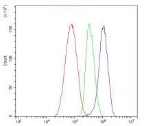
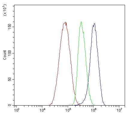
ARG58791 anti-HGD antibody FACS image
Flow Cytometry: HepG2 cells were blocked with 10% normal goat serum and then stained with ARG58791 anti-HGD antibody (blue) at 1 µg/10^6 cells for 30 min at 20°C, followed by incubation with DyLight®488 labelled secondary antibody. Isotype control antibody (green) was rabbit IgG (1 µg/10^6 cells) used under the same conditions. Unlabelled sample (red) was also used as a control.
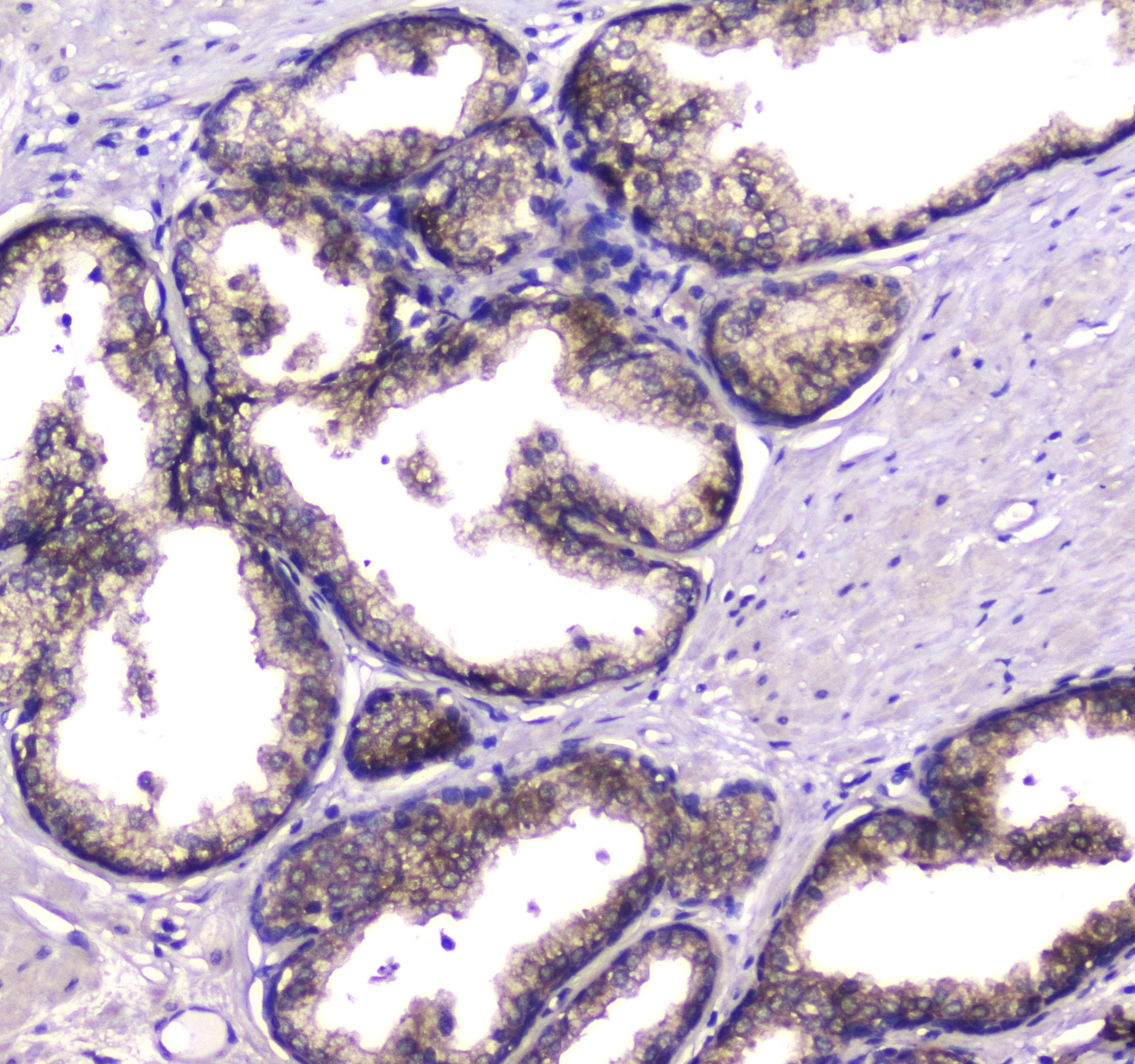
ARG58791 anti-HGD antibody IHC-P image
Immunohistochemistry: Paraffin-embedded Human prostatic cancer tissue. Antigen Retrieval: Heat mediated was performed in Citrate buffer (pH 6.0, epitope retrieval solution) for 20 min. The tissue section was blocked with 10% goat serum. The tissue section was then stained with ARG58791 anti-HGD antibody at 2 µg/ml, overnight at 4°C.


 New Products


