


anti-TXNDC5 / EndoPDI antibody


Key features and details
- 产品描述:
- 反应物种:
- 应用:
- 特异性:
- 宿主:
- 克隆:
- 同位型:
- 靶点名称:
- 抗原物种:
-
Brand:

 Product Details
| 产品描述 | Goat Polyclonal antibody recognizes TXNDC5 / EndoPDI |
|---|---|
| 反应物种 | Hu |
| 应用 | FACS, ICC/IF, IHC-P, WB |
| 特异性 | This antibody is expected to be able to recognise both reported human isoforms, as represented by NP_110437.2; NP_001139021.1. |
| 宿主 | Goat |
| 克隆 | Polyclonal |
| 同位型 | IgG |
| 靶点名称 | TXNDC5 / EndoPDI |
| 抗原物种 | Human |
| 抗原 | C-SLHRFVLSQAKDEL |
| 偶联标记 | Un-conjugated |
| 別名 | HCC2; ERp46; Thioredoxin domain-containing protein 5; HCC-2; Thioredoxin-like protein p46; UNQ364; ER protein 46; ERP46; PDIA15; ENDOPDI; STRF8; Endoplasmic reticulum resident protein 46 |
| 应用建议 |
| ||||||||||
|---|---|---|---|---|---|---|---|---|---|---|---|
| 应用说明 | WB: Recommend incubate at RT for 1h. IHC-P: Antigen Retrieval: Steam tissue section in Citrate buffer (pH 6.0). * The dilutions indicate recommended starting dilutions and the optimal dilutions or concentrations should be determined by the scientist. |
| 形式 | Liquid |
|---|---|
| 纯化 | Purified from goat serum by ammonium sulphate precipitation followed by antigen affinity chromatography using the immunizing peptide. |
| 缓冲液 | Tris saline (pH 7.3), 0.02% Sodium azide and 0.5% BSA |
| 抗菌剂 | 0.02% Sodium azide |
| 稳定剂 | 0.5% BSA |
| 浓度 | 0.5 mg/ml |
| 存放说明 | For continuous use, store undiluted antibody at 2-8°C for up to a week. For long-term storage, aliquot and store at -20°C or below. Storage in frost free freezers is not recommended. Avoid repeated freeze/thaw cycles. Suggest spin the vial prior to opening. The antibody solution should be gently mixed before use. |
| 注意事项 | For laboratory research only, not for drug, diagnostic or other use. |
| 数据库连接 | Swiss-port # Q8NBS9 Human Thioredoxin domain-containing protein 5 |
|---|---|
| 背景介绍 | This gene encodes a protein-disulfide isomerase. Its expression is induced by hypoxia and its role may be to protect hypoxic cells from apoptosis. Alternative splicing results in multiple transcript variants. Read-through transcription also exists between this gene and the neighboring upstream MUTED (muted homolog) gene. [provided by RefSeq, Dec 2010] |
| 研究领域 | Cell Biology and Cellular Response antibody; Cell Death antibody; Signaling Transduction antibody |
| 预测分子量 | 48 kDa |

ARG63498 anti-TXNDC5 / EndoPDI antibody WB image
Western blot: Human placenta lysate (RIPA buffer, 35 µg total protein per lane) stained with ARG63498 anti-TXNDC5 / EndoPDI antibody at 0.1 µg/ml dilution.

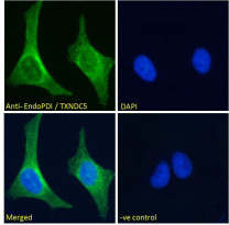
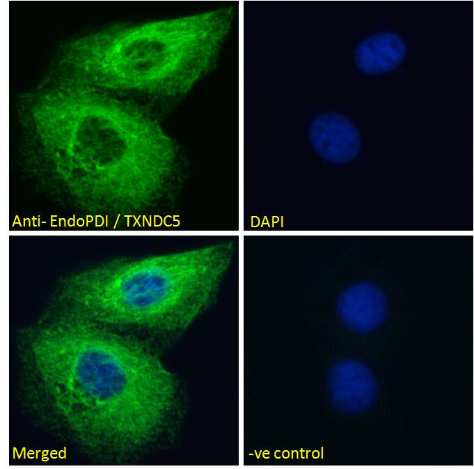
ARG63498 anti-TXNDC5 / EndoPDI antibody ICC/IF image
Immunofluorescence: Paraformaldehyde fixed HeLa cells permeabilized with 0.15% Triton. Cells were stained with ARG63498 anti-TXNDC5 / EndoPDI antibody (green) at 10 µg/ml dilution for 1 hour. DAPI (blue) for nuclear staining. Negative control: Unimmunized goat IgG (green) at 10 µg/ml dilution.
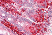
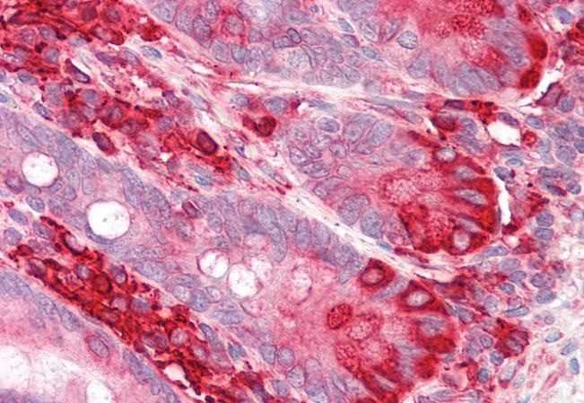
ARG63498 anti-TXNDC5 / EndoPDI antibody IHC-P image
Immunohistochemistry: Paraffin-embedded Human small intestine tissue. Antigen Retrieval: Steam tissue section in Citrate buffer (pH 6.0). The tissue section was stained with ARG63498 anti-TXNDC5 / EndoPDI antibody at 3.75 µg/ml dilution followed by AP-staining.
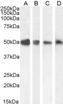
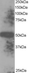
ARG63498 anti-TXNDC5 / EndoPDI antibody WB image
Western blot: 35 µg of HEK293 (A), A549 (B), HeLa (C) and HepG2 (D) cell lysates (in RIPA buffer) stained with ARG63498 anti-TXNDC5 / EndoPDI antibody at 0.1 µg/ml (A-C) and 0.01 µg/ml (D) dilutions and incubated at RT for 1 hour.
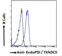
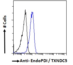
ARG63498 anti-TXNDC5 / EndoPDI antibody FACS image
Flow Cytometry: Paraformaldehyde-fixed HeLa cells permeabilized with 0.5% Triton. Cells were stained with ARG63498 anti-TXNDC5 / EndoPDI antibody (blue line) at 10 µg/ml dilution for 1 hour, followed by incubation with Alexa FluorR 488 labelled secondary antibody. IgG control: Unimmunized goat IgG (black line).
ARG63498 anti-TXNDC5 / EndoPDI antibody ICC/IF image
Immunofluorescence: Paraformaldehyde fixed U2OS cells permeabilized with 0.15% Triton. Cells were stained with ARG63498 anti-TXNDC5 / EndoPDI antibody (green) at 10 µg/ml dilution for 1 hour. DAPI (blue) for nuclear staining. Negative control: Unimmunized goat IgG (green) at 10 µg/ml dilution.


 New Products


