


anti-Visinin like 1 antibody [2D11]
![anti-Visinin like 1 antibody [2D11]](/upload/image/products/ARG52470_WB_1_191002_210_205.jpg)
![anti-Visinin like 1 antibody [2D11]](/upload/image/products/ARG52470_Visinin-like-1-2D11_WB.jpg)
![anti-Visinin like 1 antibody [2D11]](/upload/image/products/ARG52470_IHC-Fr_1_191002.jpg)
![anti-Visinin like 1 antibody [2D11]](/upload/image/products/ARG52470_IHC.JPG)
![anti-Visinin like 1 antibody [2D11]](/upload/image/products/ARG52470_WB_1_191002.jpg)
Key features and details
- 产品描述:
- 反应物种:
- 应用:
- 宿主:
- 克隆:
- 克隆号:
- 同位型:
- 靶点名称:
- 抗原物种:
-
Brand:

 Product Details
| 产品描述 | Mouse Monoclonal antibody [2D11] recognizes Visinin like 1 |
|---|---|
| 反应物种 | Hu, Ms, Rat, Bov |
| 应用 | ICC/IF, IHC-Fr, WB |
| 宿主 | Mouse |
| 克隆 | Monoclonal |
| 克隆号 | 2D11 |
| 同位型 | IgG1 |
| 靶点名称 | Visinin like 1 |
| 抗原物种 | Human |
| 抗原 | Recombinant human VSNL1 purified from E. coli |
| 偶联标记 | Un-conjugated |
| 別名 | HPCAL3; HUVISL1; Visinin-like protein 1; HLP3; Hippocalcin-like protein 3; VILIP; VLP-1; VILIP-1 |
| 应用建议 |
| ||||||||
|---|---|---|---|---|---|---|---|---|---|
| 应用说明 | Specific for the ~22k protein * The dilutions indicate recommended starting dilutions and the optimal dilutions or concentrations should be determined by the scientist. |
| 形式 | Liquid |
|---|---|
| 纯化 | Affinity Purified |
| 缓冲液 | PBS and 10 mM Sodium azide |
| 抗菌剂 | 10 mM Sodium azide |
| 存放说明 | For continuous use, store undiluted antibody at 2-8°C for up to a week. For long-term storage, aliquot and store at -20°C or below. Storage in frost free freezers is not recommended. Avoid repeated freeze/thaw cycles. Suggest spin the vial prior to opening. The antibody solution should be gently mixed before use. |
| 注意事项 | For laboratory research only, not for drug, diagnostic or other use. |
| 数据库连接 | |
|---|---|
| 基因名称 | VSNL1 |
| 全名 | visinin-like 1 |
| 背景介绍 | Visinin-like protein 1 (VSNL1), also known as VILIP1, is a calcium sensor protein expressed exclusively in neurons. Highest levels of VSNL1 expression are found in cerebellar Purkinje cells. VSNL1 has been implicated in the modulation of cell signaling cascades via regulation of adenyl cyclase activity (Braunewell et al., 1997). Additionally, VSNL1 has been associated with amyloid plaques and neurofibrillar tangles in Alzheimer’s disease (Schnurra et al., 2001). |
| 研究领域 | Cancer antibody; Signaling Transduction antibody |
| 预测分子量 | 22 kDa |
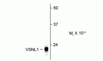
ARG52470 anti-Visinin like 1 antibody [2D11] WB image
Western blot: Rat cerebellum lysate showing specific immunolabeling of the ~ 22k VSNL1 protein stained with ARG52470 anti-Visinin like 1 antibody [2D11].

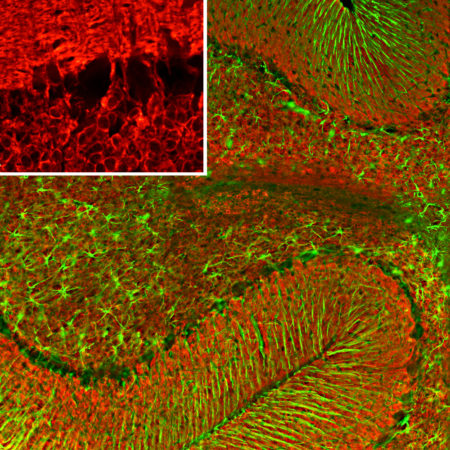
ARG52470 anti-Visinin like 1 antibody [2D11] IHC-Fr image
Immunohistochemistry: Frozen section of Rat cerebellum tissue stained with ARG52470 anti-Visinin like 1 antibody [2D11] (red) at 1:500 dilution, and costained with anti-GFAP antibody (green) at 1:5000 dilution. DAPI (blue) for nuclear staining. Following transcardial perfusion of Rat with 4% paraformaldehyde, brain was post fixed for 24 hours, cut to 45 µM, and free-floating sections were stained with the above antibodies.
Clone 2D11 reveals protein expressed in granule cell membranes and in synapses in the white matter and molecular layers of the cerebellum. The GFAP antibody stains the processes of Bergmann glia and astroglia.
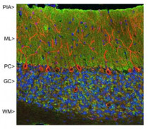
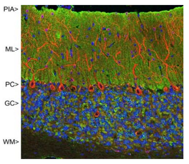
ARG52470 anti-Visinin like 1 antibody [2D11] IHC image
Immunohistochemistry: Rat cerebellum stained with ARG52470 anti-Visinin like 1 antibody [2D11] showing strong synaptic staining of VSNL1 (green) in the molecular layer (ML) and MAP2 stained with ARG52328 anti-MAP2 antibody in red.
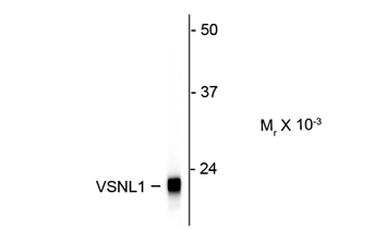
ARG52470 anti-Visinin like 1 antibody [2D11] WB image
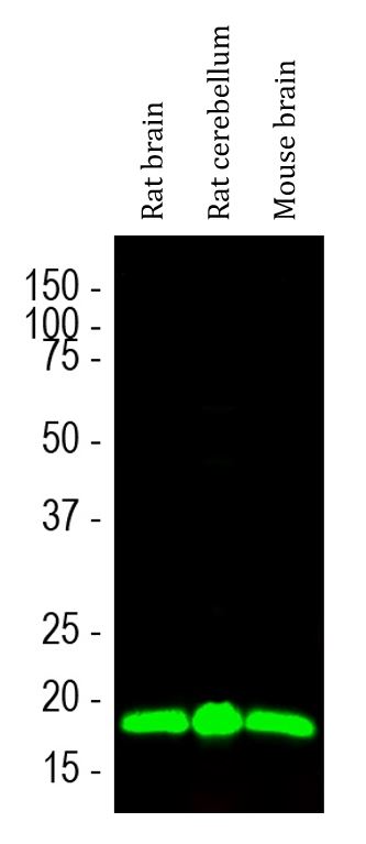
Western blot: Rat brain, Rat cerebellum and Mouse brain lysates stained with ARG52470 anti-Visinin like 1 antibody [2D11] (green) at 1:1000 dilution.
 New Products


