


Apoptosis Marker Antibody Duo (Bcl2, Bax)


Key features and details
- 产品描述:
- 靶点名称:
- 別名:
-
Brand:

 Product Details
| 货号 | 内含物名称 | 宿主克隆性 | 反应 | 应用 | 包装 |
|---|---|---|---|---|---|
| ARG65612 | anti-Bax antibody | Rabbit pAb | Hu, Ms, Rat | ICC/IF, IHC-P, WB | 50 μl |
| ARG55188 | anti-Bcl 2 antibody | Rabbit pAb | Hu, Ms, Rat | ICC/IF, IP, WB | 50 μl |
| 产品描述 | BCL2 suppresses apoptosis in a variety of cell systems including factor-dependent lymphohematopoietic and neural cells. Regulates cell death by controlling the mitochondrial membrane permeability. Appears to function in a feedback loop system with caspases. Inhibits caspase activity either by preventing the release of cytochrome c from the mitochondria and/or by binding to the apoptosis-activating factor (APAF-1). BCL2 family members form hetero- or homodimers and act as anti- or pro-apoptotic regulators that are involved in a wide variety of cellular activities.Bax protein belongs to the BCL2 protein family. This protein forms a heterodimer with BCL2, and functions as an apoptotic activator. This protein is reported to interact with, and increase the opening of, the mitochondrial voltage-dependent anion channel (VDAC), which leads to the loss in membrane potential and the release of cytochrome c. (provided by RefSeq, Jul 2008)BAX/BCL2 expression ratio has been identified as an import index to figure out the fate of the cell undergoing apoptosis or anti-apoptosis in variety studies, including cancer study, therapeutic agent searching and therapeutic response evaluation.arigo provide an Apoptosis marker Duo, ARG30268, including anti-Bcl2 and anti-Bax antibodies, is useful for user in studying BAX/BCL2 correlation. |
|---|---|
| 靶点名称 | Apoptosis Marker |
| 別名 | Apoptosis Marker antibody; Bcl 2 antibody; Bax antibody |
| 存放说明 | For continuous use, store undiluted antibody at 2-8°C for up to a week. For long-term storage, aliquot and store at -20°C or below. Storage in frost free freezers is not recommended. Avoid repeated freeze/thaw cycles. Suggest spin the vial prior to opening. The antibody solution should be gently mixed before use. |
|---|---|
| 注意事项 | For laboratory research only, not for drug, diagnostic or other use. |
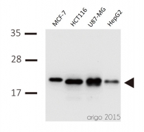
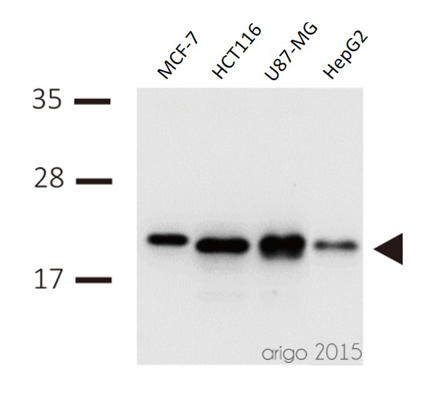
ARG65612 anti-Bax antibody WB image
Western blot: 30 µg of MCF-7, HCT116, U87-MG, and HepG2 cell line lysates stained with ARG65612 anti-Bax antibody at 1:500 dilution.
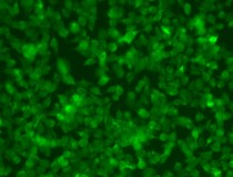
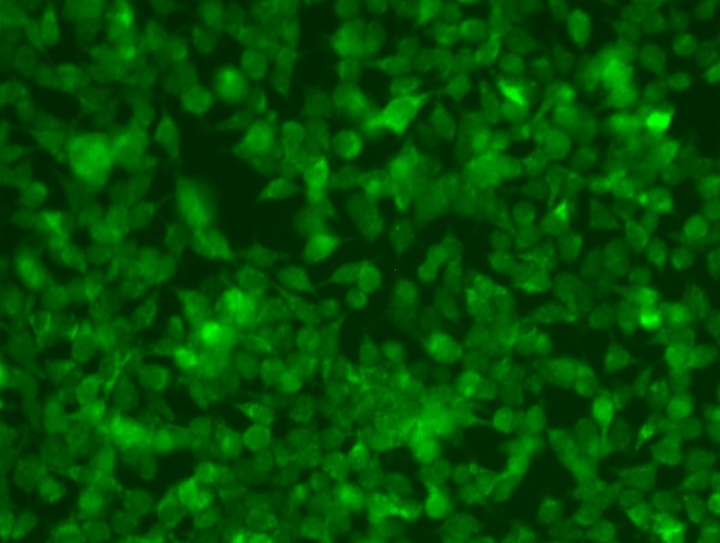
ARG65612 anti-Bax antibody ICC/IF image
Immunocytochemistry: HeLa cells fixed with Methanol / Acetone 1:1 ratio (-20°C for 20 min) and blocked by 3% BSA in PBS (RT for 1 hour). The cells stained with ARG65612 anti-Bax antibody at 1:20 dilution (RT for 1 hour).

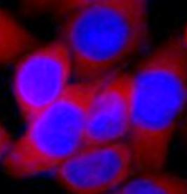
ARG55188 anti-Bcl 2 antibody ICC/IF image
Immunofluorescence: HeLa cells stained with ARG55188 anti-Bcl 2 antibody (red) at 1:100 dilution. DAPI (blue) for nuclear staining.
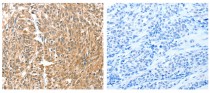
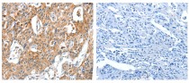
ARG65612 anti-Bax antibody IHC-P image
Immunohistochemistry: paraffin-embedded Human esophagus cancer tissue stained with ARG65612 anti-Bax antibody (left) at 1/30 dilution, or the same antibody preincubated with antigen (right). (Original magnification: X200)
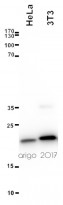
ARG65612 anti-Bax antibody WB image
Western blot: 20 µg of HeLa and 3T3 cell lysates stained with ARG65612 anti-Bax antibody at 1:3000 dilution.
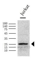
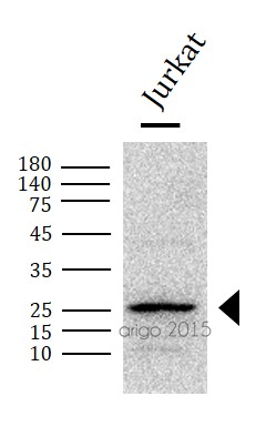
ARG55188 anti-Bcl-2 antibody WB image
Western blot: 30 μg of Jurkat cell lysate stained with ARG55188 anti-Bcl-2 antibody at 1:1000 dilution.
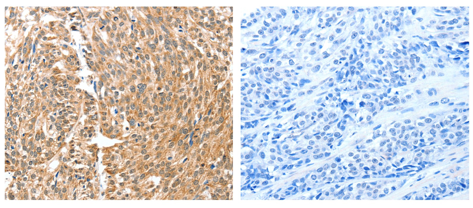
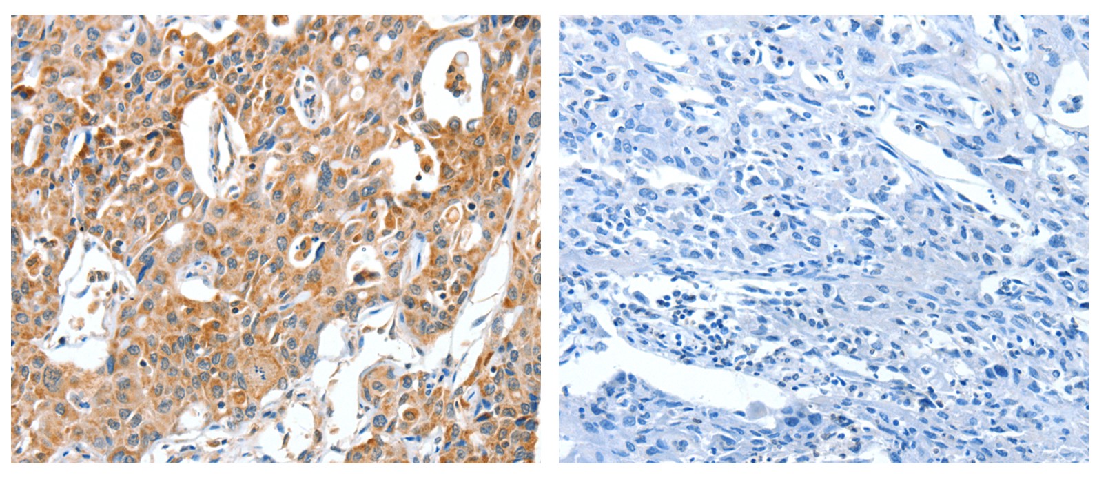
ARG65612 anti-Bax antibody IHC-P image
Immunohistochemistry: paraffin-embedded Human lung cancer tissue stained with ARG65612 anti-Bax antibody (left) at 1/30 dilution, or the same antibody preincubated with antigen (right). (Original magnification: X200)


Transcutaneous electrical acupoint stimulation alleviates cerebral ischemic injury through the TLR4/MyD88/NF-κ B pathway
ARG55188: WB / Rat


Suppression of gastric cancer cell proliferation by miR-494-3p inhibitor-loaded engineered exosomes
ARG55188: WB / Human
Astaxanthin alleviates fine particulate matter (PM2.5)-induced lung injury in rats by suppressing ferroptosis and apoptosis
ARG55188: WB / Rat
Probucol attenuates high glucose-induced Müller cell damage through enhancing the Nrf2/p62 signaling pathway
ARG55188: WB / Human
Dapagliflozin protects against doxorubicin-induced nephrotoxicity associated with nitric oxide pathway-A translational study
ARG55188: WB / Rat
Honokiol attenuates mitochondrial fission and cell apoptosis by activating Sirt3 in intracerebral hemorrhage
ARG55188: WB / Rat
Bone targeted miRNA delivery system for miR-34a with enhanced anti-tumor efficacy to bone-associated metastatic breast cancer
ARG55188: WB / Human
Resveratrol improves ovarian state by inhibiting apoptosis of granulosa cells
ARG55188: WB / Human
Dapagliflozin-entresto protected kidney from renal hypertension via downregulating cell-stress signaling and upregulating SIRT1/PGC-1α/Mfn2-medicated mitochondrial homeostasis
ARG55188: WB / Rat
Ivabradine could not decrease mitral regurgitation triggered atrial fibrosis and fibrillation compared with carvedilol
ARG55188: WB / Rat
Transpulmonary Expression of Exosomal microRNAs in Idiopathic and Congenital Heart Disease-Related Pulmonary Arterial Hypertension
ARG55188: WB / Human
Preclinical Therapeutic Assessment of a New Chemotherapeutics [Dichloro(4,4'-Bis(2,2,3,3-Tetrafluoropropoxy) Methyl)-2,2'-Bipryridine) Platinum] in an Orthotopic Patient-Derived Xenograft Model of Triple-Negative Breast Cancers
ARG55188, ARG65612: WB / Human
Orlistat Resensitizes Sorafenib-Resistance in Hepatocellular Carcinoma Cells through Modulating Metabolism
ARG55188:WB / Human
Deletion of MicroRNA-21 Impairs Neovascularization Following Limb Ischemia: From Bedside to Bench
ARG55188: WB / Mouse
Cobalt protoporphyrin promotes human keratinocyte migration under hyperglycemic conditions
ARG65612: WB / Human
Naringin Interferes Doxorubicin-Induced Myocardial Injury by Promoting the Expression of ECHS1
ARG55188: WB / Rat
Kansuinine A Ameliorates Atherosclerosis and Human Aortic Endothelial Cell Apoptosis by Inhibiting Reactive Oxygen Species Production and Suppressing IKKβ/IκBα/NF-κB Signaling
ARG65612: WB / Human
Circ_0081572 inhibits the progression of periodontitis through regulating the miR-378h/RORA axis.
ARG55188: WB / Human
Dapagliflozin Improves Cardiac Hemodynamics and Mitigates Arrhythmogenesis in Mitral Regurgitation-Induced Myocardial Dysfunction.
ARG55188: WB / Rat
miR-371b-5p promotes cell proliferation, migration and invasion in non-small cell lung cancer via SCAI
ARG55188: WB / Human
Xenogeneic and Allogeneic Mesenchymal Stem Cells Effectively Protect the Lung Against Ischemia-reperfusion Injury Through Downregulating the Inflammatory, Oxidative Stress, and Autophagic Signaling Pathways in Rat.
ARG55188: WB / Human
Multi-omics analysis of pathological changes in the amygdala of rats subjected to chronic restraint stress.
ARG65612: WB / Rat
Possible Involvement of PTEN Signaling Pathway in the Anti-apoptotic Effect of Electroacupuncture Following Ischemic Stroke in Rats
ARG55188: WB / Rat
Electroacupuncture Alleviated Neuronal Apoptosis Following Ischemic Stroke in Rats via Midkine and ERK/JNK/p38 Signaling Pathway.
ARG55188: WB / Rat
A LY-15, a novel cyclic pentapeptide that inhibits B16 cell proliferation and migration and induces cell apoptosis
ARG55188, ARG65612: WB / Mouse
New Products


