


BCL-2 Rabbit Monoclonal Antibody(ARA941)

Key features and details
- Reactivity:Human
- Application:IHC
- Host:Rabbit
- Clonality:Monoclonal
- Target:BCL-2
-
Brand:

CAT.NO. : ARA6782
RMB Please choose
RMB Please choose
*产品价格可能会有所调整,请以品牌方官网实时更新的价格为准,以确保准确性。
 Product Details
Background
Proteins of the Bcl-2 family are regulators of apoptosis (programmed cell death) localized to membranes of primarily mitochondria, but also to smooth endoplasmic reticulum and nucleolemma. The fine balance between pro- and anti-apoptotic Bcl-2 family members regulates the cell fate in response to many signalling pathways. Altered expression of the proteins may lead to either premature cell death or to inappropriate cell survival promoting neoplastic growth. Bcl-2, Bax and Bcl-X are the most well known proteins in this family. In neoplastic lesions Bcl-2 upregulation may act by suppression of programmed cell death and extension of the tumour cell life span. Bcl-2 overexpression contributes to increased resistance to chemotherapy. However, the prognostic value of Bcl-2 overexpression is dependent on the tumour type, and in some types BCl-2 may even have a tumour suppressor effect. Overexpression of Bcl-2 is common in many types of cancer, including non-Hodgkin's lymphoma and leukaemias, adenocarcinomas (e.g., prostate, colorectum, stomach, and lung), squamous cell carcinoma, small cell carcinoma, neuroblastoma and various sarcomas. Bcl-2 is helpful in distinguishing follicular lymphoma from reactive follicular hyperplasia. Bcl-2 staining can not be used for differential diagnosis between follicular lymphoma and other types of low-grade B-cell lymphomas since most of the latter also express bcl-2. Monocytoid B-cell hyperplasia contrasts with bcl-2 negativity against positive reaction in 80% of the marginal zone lymphomas. Possibly, Bcl-2 may aid in the differential diagnosis of sarcomas. Tonsil is recommendable as positive and negative tissue control for BCL2. A moderate to strong, predominantly cytoplasmic staining reaction should be displayed in virtually all T-cells and B-cells in the mantle zone of the reactive follicles, whereas the majority of the basal squamous epithelial cells, e.g. lining the tonsillar crypts, must show an at least weak staining intensity. Germinal centre T-cells should be distinctively demonstrated, whereas germinal centre B-cells should be negative.
Application
|
Application |
Dilution Ratio |
|
IHC-P |
1:1,000-1:5,000 |
Overview
|
Antibody Type |
Recombinant Rabbit Monoclonal Antibody |
|
Immunogen |
Synthetic peptide within Human Bcl2 alpha aa 50 - 150 (N terminal). |
|
Species Reactivity |
Human |
|
Validated Applications |
WB, IHC - P, IF - Cell, FC |
|
Molecular Weight |
Predicted band size: 26 kDa |
|
Positive Control |
MCF7 cell lysate, Jurkat cell lysate, HL - 60 cell lysate, THP - 1 cell lysate, human B - cell lymphoma tissue, human tonsil tissue, human lymph nodes tissue, HeLa, THP - 1 |
|
Conjugation |
unconjugated |
|
Form |
Liquid |
|
Concentration |
1μg/ul |
|
Storage Buffer |
PBS (pH7.4), 0.1% BSA, 40% Glycerol. Preservative: 0.05% Sodium Azide |
|
Isotype |
IgG |
|
Purification Method |
Protein A affinity purified |
Data
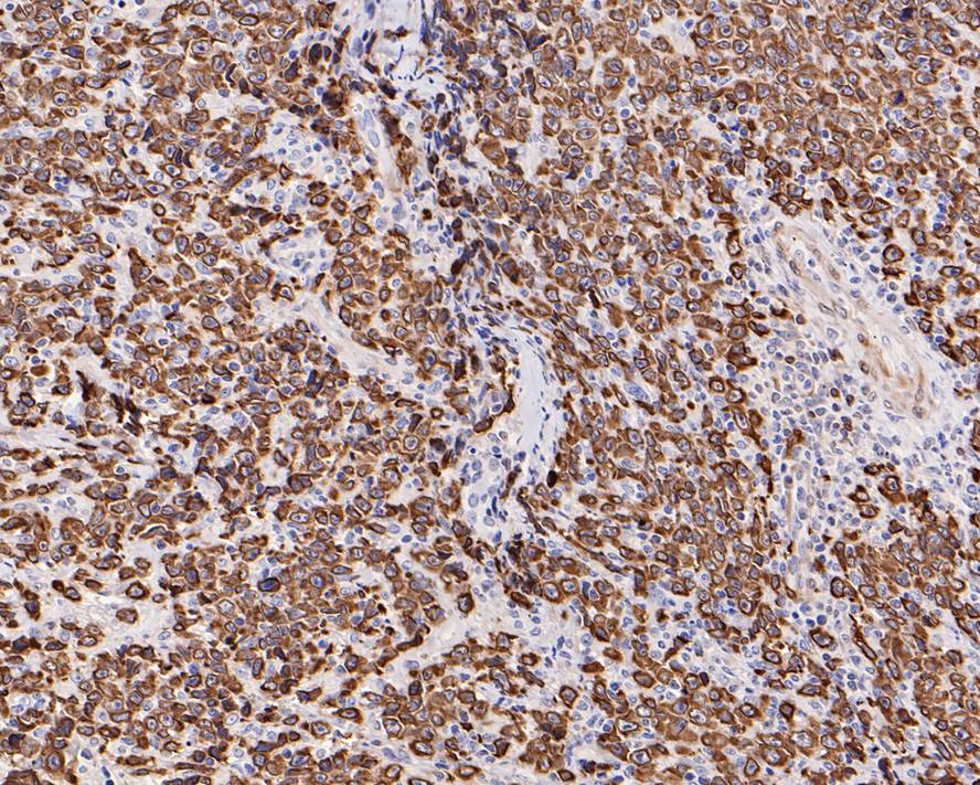
Immunohistochemical staining of human B-cell lymphoma tissue using BCL-2 Rabbit Monoclonal Antibody(ARA941)
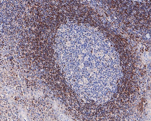
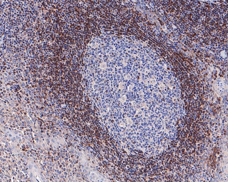
Immunohistochemical staining of human tonsil tissue using BCL-2 Rabbit Monoclonal Antibody(ARA941)
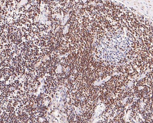
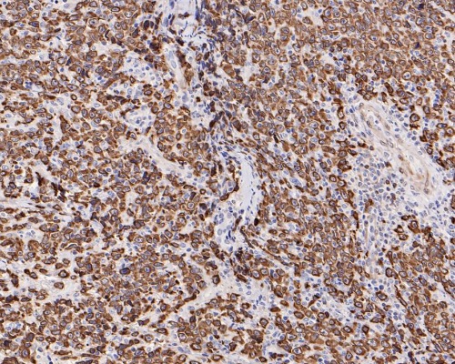
Immunohistochemical staining of human lymph nodes tissue using BCL-2 Rabbit Monoclonal Antibody(ARA941)
Storage
Store at 4°C short term. For long term storage, store at -20°C, avoiding freeze/thaw cycles.
Research Use Only
For Research Use Only. Not for use in diagnostic procedures.
New Products


