


Claudin 1 Rabbit polyclonal antibody


Key features and details
- Reactivity:Human, Mouse, Rat,Bovine,Dog,Sheep
- Application:WB, IHC, IF/ICC
- Host:Rabbit
- Clonality: polyclonal
- Target:Claudin 1
-
Brand:

CAT.NO. : ARA6613
RMB Please choose
RMB Please choose
*产品价格可能会有所调整,请以品牌方官网实时更新的价格为准,以确保准确性。
 Product Details
Background
Claudin-1 is a member of the transmembrane protein family claudins located in cell-cell tight junctions and it acts as a co-receptor for HCV entry into hepatic cells. Claudins are abundant in luminal epithelial sheets where they maintain epithelial cell polarity. Claudin-1 is expressed in most tissues such as bladder, fallopian tube, liver, pancreas, prostate, and skin.
Application
|
Applications |
Dilution |
|
WB |
1:500 - 1:1000 |
|
IH |
1:50 - 1:100 |
|
IF/IC |
1:50 - 1:200 |
Overview
|
Purification Method |
The antibody was purified by immunogen affinity chromatography. |
|
Clonality |
Polyclonal |
|
Product Form |
Liquid in 0.42% Potassium phosphate, 0.87% Sodium chloride, pH 7.3, 30% glycerol, and 0.01% sodium azide. |
|
Gene Name |
CLDN1 |
|
Related Names |
CLD1; SEMP1; Claudin-1; Senescence-associated epithelial membrane protein |
|
Gene ID (Human) |
9076 |
|
Gene ID (Mouse) |
12737 |
|
Gene ID (Rat) |
65129 |
|
Protein ID (Human) |
O95832 |
|
Protein ID (Mouse) |
O88551 |
|
Protein ID (Rat) |
P56745 |
Data
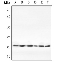
Western blot analysis of Claudin 1 expression in Hela (A), H1688 (B), mouse liver (C), mouse kidney (D), rat liver (E), rat kidney (F) whole cell lysates. (Predicted band size: 22 kD; Observed band size: 22 kD)

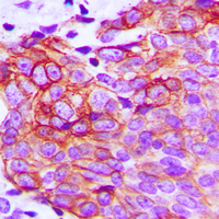
Immunohistochemical analysis of Claudin 1 staining in human breast cancer formalin fixed paraffin embedded tissue section. The section was pre-treated using heat mediated antigen retrieval with sodium citrate buffer (pH 6.0). The section was then incubated with the antibody at room temperature and detected using an HRP conjugated compact polymer system. DAB was used as the chromogen. The section was then counterstained with haematoxylin and mounted with DPX.
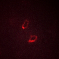
Immunofluorescent analysis of Claudin 1 staining in HeLa cells. Formalin-fixed cells were permeabilized with 0.1% Triton X-100 in TBS for 5-10 minutes and blocked with 3% BSA-PBS for 30 minutes at room temperature. Cells were probed with the primary antibody in 3% BSA-PBS and incubated overnight at 4 °C in a hidified chamber. Cells were washed with PBST and incubated with a AF594-conjugated secondary antibody (red) in PBS at room temperature in the dark.
Storage
Store at 4°C short term. For long term storage, store at -20°C, avoiding freeze/thaw cycles.
New Products


