


eNOS Rabbit Polyclonal Antibody

Key features and details
- Reactivity:Human,Mouse,Rat,Bovine,Dog,Rabbit,Sheep
- Application:WB,IHC,IF/ICC
- Host:Rabbit
- Clonality:Polyclonal
- Target:eNOS
-
Brand:

 Product Details
Nitric oxide (NO) is an inorganic, gaseous free radical that carries a variety of messages between cells. Vasorelaxation, neurotransmission and cytotoxicity can all be potentiated through cellular response to NO. NO production is mediated by members of the nitric oxide synthase (NOS) family. NOS catalyzes the oxidization of L-arginine to produce L-citrulline and NO. Two constitutive isoforms, brain or neuronal NOS (b or nNOS, type I) and endothelial cell NOS (eNOS, type III), and one inducible isoform (iNOS, type II), have been cloned. All NOS isoforms contain calmodulin, nicotinamide adenine dinucleotide phosphate (NADPH), flavin adenine dinucleotide (FAD), and flavin mononucleotide (FMN) binding domains.
|
Applications |
Tested Dilution |
|
WB |
1:500 - 1:1000 |
|
IHC |
1:100 - 1:200 |
|
IF/IC |
1:100 - 1:500 |
|
Immunogen |
KLH - conjugated synthetic peptide encompassing a sequence within the C - term region of human eNOS. The exact sequence is proprietary. |
|
Purification Method |
The antibody was purified by immunogen affinity chromatography. |
|
Clonality |
Polyclonal |
|
Product Form |
Liquid in 0.42% Potassium phosphate, 0.87% Sodium chloride, pH 7.3, 30% glycerol, and 0.01% sodium azide. |
|
Dilution Ratios |
WB (1/500 - 1/1000), IH (1/100 - 1/200), IF/IC (1/100 - 1/500) |
|
Gene Name |
NOS3 |
|
Related Names |
Nitric oxide synthase, endothelial; Constitutive NOS; cNOS; EC - NOS; Endothelial NOS; eNOS; NOS type III; NOSIII |
|
Gene ID (Human) |
4846 |
|
Gene ID (Mouse) |
18127 |
|
Gene ID (Rat) |
24600 |
|
Protein ID (Human) |
P29474 |
|
Protein ID (Mouse) |
P70313 |
|
Protein ID (Rat) |
Q62600 |
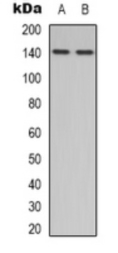
Western blot analysis of eNOS expression in HepG2 (A), Hela (B) whole cell lysates. (Predicted band size: 133; 69 kD; Observed band size: 140 kD)

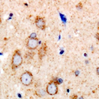
Immunohistochemical analysis of eNOS staining in human brain formalin fixed paraffin embedded tissue section. The section was pre-treated using heat mediated antigen retrieval with sodium citrate buffer (pH 6.0). The section was then incubated with the antibody at room temperature and detected using an HRP conjugated compact polymer system. DAB was used as the chromogen. The section was then counterstained with haematoxylin and mounted with DPX.

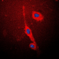
Immunofluorescent analysis of eNOS staining in MCF7 cells. Formalin-fixed cells were permeabilized with 0.1% Triton X-100 in TBS for 5-10 minutes and blocked with 3% BSA-PBS for 30 minutes at room temperature. Cells were probed with the primary antibody in 3% BSA-PBS and incubated overnight at 4 °C in a hidified chamber. Cells were washed with PBST and incubated with a DyLight 594-conjugated secondary antibody (red) in PBS at room temperature in the dark. DAPI was used to stain the cell nuclei (blue).
New Products


