


GFAP Rabbit Monoclonal Antibody(ARB959)

Key features and details
- Target:
- Clone ID:
- Host:
- Molecular Weight:
- Purity:
- Species Cross-reactivity:
- Applications:
- Swissprot ID:
-
Brand:

CAT.NO. : ARB6751
RMB Please choose
RMB Please choose
Size:
Trail, Bulk size or Custom requests Please contact us
*产品价格可能会有所调整,请以品牌方官网实时更新的价格为准,以确保准确性。
 Product Details
Background
GFAP (Glial fibrillary acidic protein) is an intermediate filament protein. The protein is the smallest (8 nm) of the intermediate filament proteins with a molecular weight of about 50 kDa.
In the central nervous system, GFAP is expressed in astrocytes and ependymal cells but not in other glial cells. However, immature oligodendrocytes and immature choroid plexus cells may be GFAP positive. In the peripheral nervous system enteric Schwann cells and satellite cells of human sensory ganglia express GFAP. Outside the nervous system, GFAP is seen in myoepithelial cells and chondroblasts. In tumor tissues, astrocytoma, ependymoma, glioblastoma and oligodendroglioma are almost always positive. Plexus carcinoma, ganglioglioma and primitive neuroectodermal tumors (PNET: neuroblastoma a.o.) express GFAP to a varying extent. Schwannoma and neurofibroma frequently express GFAP. Chondroma, chondrosarcoma and pleomorphic adenoma are also GFAP positive in most cases. A few carcinomas (especially from lung and breast) may express GFAP in paraganglioma GFAP may be detected in sustentacular cells.
GFAP is used to differentiate astrocytoma from non-glial cell tumors.
Application
|
Application |
Dilution Ratio |
|
IHC |
1:100 - 1:200 |
Overview
|
Predicted Molecular Wt |
50kDa |
|
Species Cross-reactivity |
Human |
|
Applications |
IHC-P |
|
Purity |
ProA affinity purified IgG |
|
Form |
Liquid |
|
Swissprot ID |
P14136 |
|
Subcellular location |
Cytoplasm |
|
Recommended method |
Heat induced epitope retrieval with Tris-EDTA buffer (pH 9.0), primary antibody incubate at RT (18℃-25℃) for 30 minutes |
|
Immunogen |
Synthetic peptide corresponding to GFAP residues within aa332-432 of GFAP was used as an immunogen |
|
Storage Buffer |
PBS 59%, Sodium azide 0.01%, Glycerol 40%, BSA 0.05% |
Data
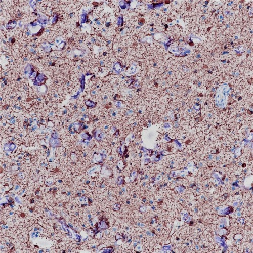
Immunohistochemical staining of human brain tissue using GFAP Rabbit Monoclonal Antibody(ARB959)
Storage
Store at -20°C. Stable for one year from the date of shipment.
Research Use Only
For Research Use Only. Not for use in diagnostic procedures.
New Products


