


GGT1 Mouse Monoclonal Antibody (F-7)
Key features and details
- Reactivity:Mouse, Rat
- Application:WB, IP, IF, IHC(P) , ELISA
- Host:Mouse
- Clonality:Monoclonal
- lsotype:IgG2b
- Target Name:GGT1
-
Brand:

CAT.NO. : ARA0107
RMB Please choose
RMB Please choose
Size:
Trail, Bulk size or Custom requests Please contact us
*产品价格可能会有所调整,请以品牌方官网实时更新的价格为准,以确保准确性。
 Product Details
BACKGROUND
GGT (γ - glutamyltranspeptidase) acts as a glutathionase and catalyzes the transfer of the glutamyl moiety of glutathione to a variety of amino acids and dipeptide acceptors. This enzyme is located on the outer surface of the cell membrane and is widely distributed in mammalian tissues involved in absorption and secretion. In humans, hepatic GGT activity is elevated in some liver diseases. GGT1 is released into the bloodstream after liver damage, and an elevated level of the enzyme may be a useful early sign of hepatocellular carcinoma. GGT5 converts leukotriene C4 to leukotriene D4; it does not, however, convert synthetic substrates that are commonly used to assay GGT. In human serum and in human tissues, there is a marked heterogeneity in GGT, but this heterogeneity can be attributed to different glycosylation of the same peptide rather than to the products of different genes.
Application
apical mem is recommended for detection of GGT1 heavy chain of mouse and rat origin by Western Blotting (starting dilution 1:100, dilution range 1:100 - 1:1000), immunoprecipitation [1 - 2 µg per 100 - 500 µg of total protein (1 ml of cell lysate)], immunofluorescence (starting dilution 1:50, dilution range 1:50 - 1:500), immunohistochemistry (including paraffin - embedded sections) (starting dilution 1:50, dilution range 1:50 - 1:500) and solid phase ELISA (starting dilution 1:30, dilution range 1:30 - 1:3000).
Data
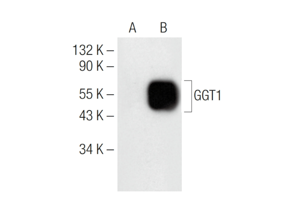
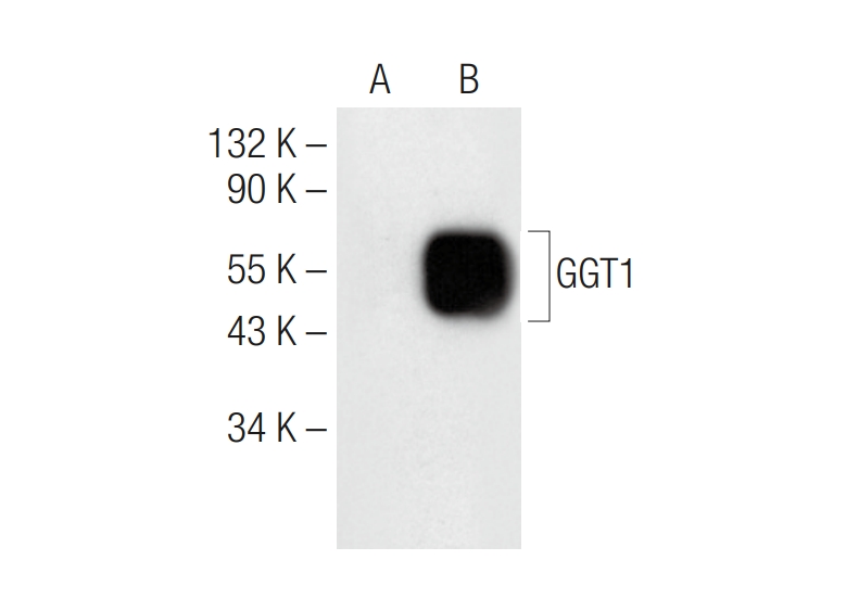
GGT1 (F - 7). Western blot analysis of GGT1 expression in non - transfected (A) and mouse GGT1 transfected (B) 293T whole cell lysates.


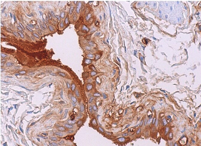
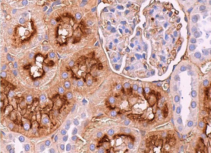
GGT1 (F - 7). Immunoperoxidase staining of formalin fixed, paraffin - embedded rat urinary bladder tissue showing cytoplasmic staining of urothelial cells (above). Immunoperoxidase staining of formalin fixed, paraffin - embedded rat kidney tissue showing apical membrane and cytoplasmic staining of cells in tubules (below).
New Products


