Intermediate Neurofilament Antibody Panel (NF-L, NF-M,NF-H)


Key features and details
- 产品描述:
- 靶点名称:
- 別名:
-
Brand:

 Product Details
| 货号 | 内含物名称 | 宿主克隆性 | 反应 | 应用 | 包装 |
|---|---|---|---|---|---|
| ARG52348 | anti-Neurofilament NF-L antibody [DA2] | Mouse mAb | Hu, Ms, Rat | FACS, ICC/IF, IHC-Fr, IHC-P, WB | 50 μl |
| ARG52351 | anti-Neurofilament NF-M antibody | Chicken pAb | Ms, Rat | ICC/IF, IHC-Fr, WB | 50 μl |
| ARG52347 | anti-Neurofilament NF-H antibody | Chicken pAb | Ms, Rat, Cow | ICC/IF, IHC-Fr, WB | 50 μl |
| ARG65350 | Goat anti-Mouse IgG antibody (HRP) | Goat pAb | Ms | ELISA, IHC-P, WB | 50 μl |
| 产品描述 | Neurofilaments are the major intermediate filaments found in neurons and consist of light (NFL), medium (NFM), and heavy (NFH) subunits. It appears that the essence of this assembly process is the heterotetrameric unit, which exists in two forms: NFL-NFM and NFL-NFH. Tetramer pairs form protofilaments and eight protofilaments constitute the final 10 nm intermediate filament. NFL is essential for the correct assembly of neurofilaments and maintenance of axonal calibre and NFM not only forms cross-bridges, stabilizing the filament network, but also helps in longitudinal extension. NFH also forms cross-bridges and may interact with micro-tubules and other cytoskeletal elements. Neurofilament accumulations are found in several human neurological diseases. These include ALS, Lewy bodies in Parkinson’s disease and some dementias (neurofilaments are a component of the Lewy body along with a-synuclein), Alzheimer’s disease, progressive supranuclear palsy, Charcot-Marie-Tooth disease, diabetic neuropathy, and giant axonal neuropathy. Cohlberg, J.A. et al. (1995) J. Biol. Chem. 270, 9334-9339. Sakaguchi, T et al. (1993) Neurosci Lett 153, 65–68. Ammar Al-Chalabi and Christopher C. J. Miller (2003) BioEssays 25, 346–355. |
|---|---|
| 靶点名称 | Intermediate Neurofilament |
| 別名 | Intermediate Neurofilament antibody; Neurofilament NF-H antibody; Neurofilament NF-L antibody; Neurofilament NF-M antibody |
| 存放说明 | For continuous use, store undiluted antibody at 2-8°C for up to a week. For long-term storage, aliquot and store at -20°C or below. Storage in frost free freezers is not recommended. Avoid repeated freeze/thaw cycles. Suggest spin the vial prior to opening. The antibody solution should be gently mixed before use. |
|---|---|
| 注意事项 | For laboratory research only, not for drug, diagnostic or other use. |
| 全名 | Antibody Panel for Intermediate Neurofilament (NF-L, NF-M,NF-H) |
|---|---|
| 研究领域 | Controls and Markers antibody; Developmental Biology antibody; Immune System antibody; Neuroscience antibody; Signaling Transduction antibody |
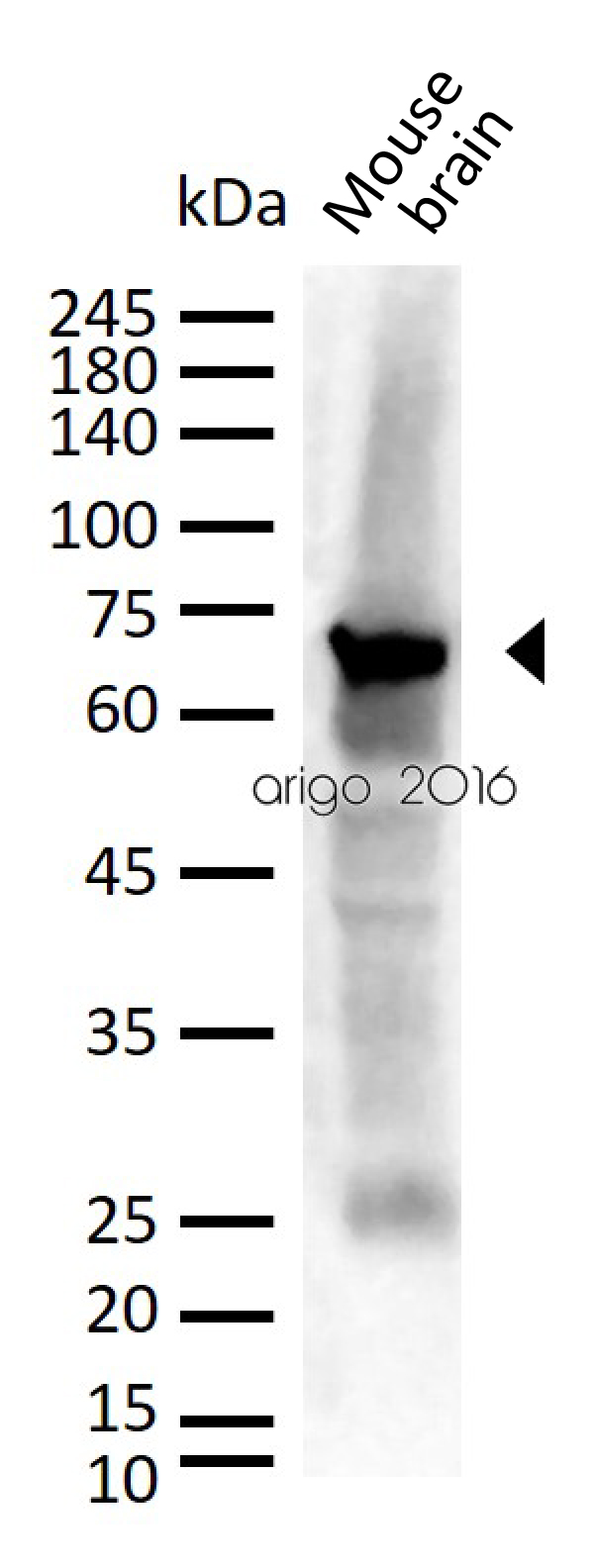
ARG52348 anti-Neurofilament NF-L antibody [DA2] WB image
Western blot: 30 µg of Mouse brain lysate stained with ARG52348 anti-Neurofilament NF-L antibody [DA2] at 1:1000 dilution.
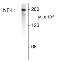
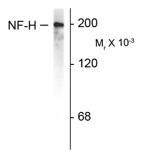
ARG52347 anti-Neurofilament NF-H antibody WB image
Western blot: rat cortex lysate showing specific immunolableing of the ~200k NF-H protein stained with ARG52347 anti-Neurofilament NF-H antibody.

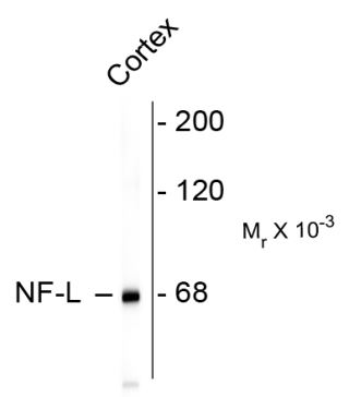
ARG52348 anti-Neurofilament NF-L antibody [DA2] WB image
Western blot: rat cortex lysate stained with ARG52348 anti-Neurofilament NF-L antibody [DA2] showing specific immunolableing of the ~ 68k NF-L protein.
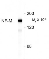
ARG52351 anti-Neurofilament NF-M antibody WB image
Western blot: rat cortex lysate stained with ARG52351 anti-Neurofilament NF-M antibody showing specific immunolabeling of the ~ 145k NF-M protein.

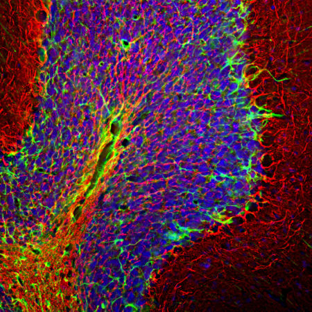
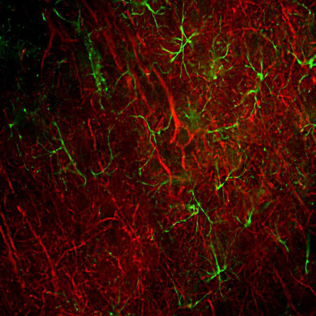
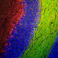
ARG52347 anti-Neurofilament NF-H antibody IHC-Fr image
Immunohistochemistry: Frozen section of Rat cerebellum tissue stained with ARG52347 anti-Neurofilament NF-H antibody (red) at 1:5000 dilution, and costained with anti-GFAP antibody (green) at 1:5000 dilution. DAPI (blue) for nuclear staining. Following transcardial perfusion with 4% paraformaldehyde, brain was post fixed for 24 hours, cut to 45 µM, and free floating sections were stained with above antibodies.
The NF-H antibody labels network of axons of different neurons, while the GFAP antibody stains astrocytes and other glial cells.

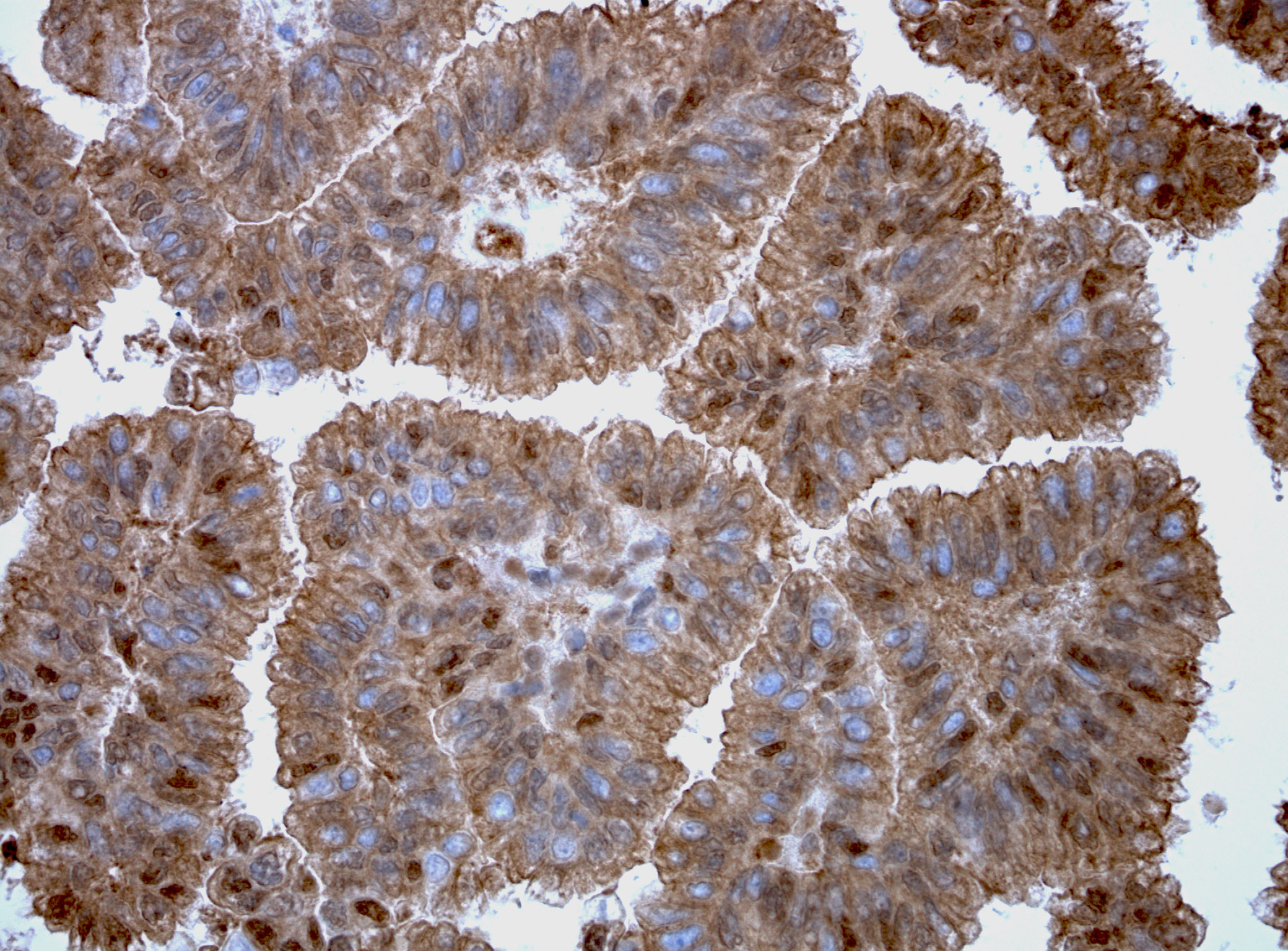
ARG52348 anti-Neurofilament NF-L antibody [DA2] IHC-Fr image
Immunohistochemistry: Frozen section of Rat frontal cortex tissue stained with ARG52348 anti-Neurofilament NF-L antibody [DA2] (red) at 1:500 dilution, and costained with anti-GFAP antibody (green) at 1:5000 dilution. Following transcardial perfusion of Rat with 4% paraformaldehyde, brain was post fixed for 24 hours, cut to 45 µM, and free-floating sections were stained with above antibodies.
Clone DA2 labels cell bodies and processes of pyramidal neurons, as well as dendrites and axons of other neuronal cells, while the GFAP antibody stains the network of glial cells.
ARG52351 anti-Neurofilament NF-M antibody IHC-Fr image
Immunohistochemistry: Frozen section of Rat cerebellum tissue stained with ARG52351 anti-Neurofilament NF-M antibody (red) at 1:1000 dilution, and co-stained with ARG11063 anti-CNPase antibody [1H10] (green) at 1:500 dilution. DAPI (blue) for nuclear staining. (Sample preparation: Following transcardial perfusion of rat with 4% paraformaldehyde, brain was post fixed for 24 hours, cut to 45 µM, and free-floating sections were stained with the above antibodies.)
The NF-M antibody labels the network of axons of basket neurons and other neurons. The CNPase antibody stains oligodendrocytes, cells that create myelin sheaths around axons.
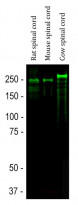
ARG52347 anti-Neurofilament NF-H antibody WB image
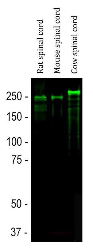
Western blot: Rat spinal cord, Mouse spinal cord and Cow spinal cord lysates stained with ARG52347 anti-Neurofilament NF-H antibody (green) at 1:20000 dilution.
Strong band at about 200-220 kDa corresponds to the phosphorylated from of NF-H. Smaller proteolytic fragments of NF-H are also detected in spinal cord preparations with this antibody.
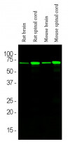
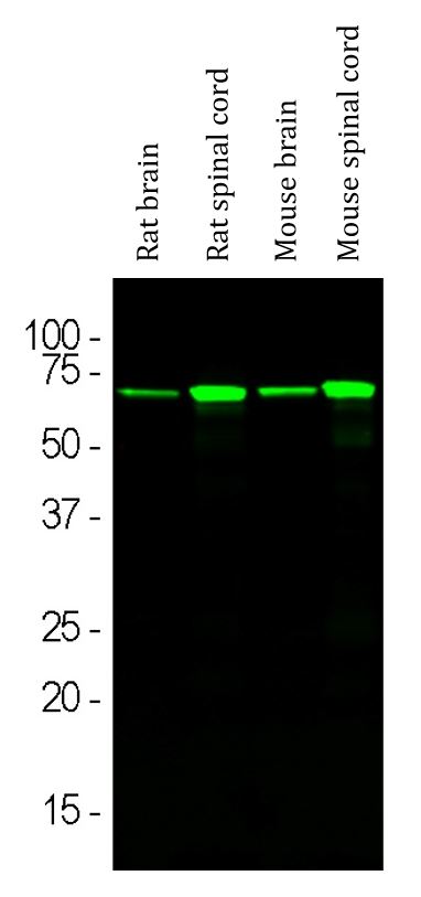
ARG52348 anti-Neurofilament NF-L antibody [DA2] WB image
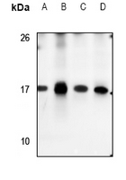
Western blot: Rat brain, Rat spinal cord, Mouse brain and Mouse spinal cord lysates stained with ARG52348 anti-Neurofilament NF-L antibody [DA2] (green) at 1:5000 dilution.
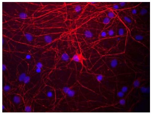
ARG52347 anti-Neurofilament NF-H antibody ICC/IF image
Immunofluorescence: rat cortical neurons and glia stained with ARG52347 anti-Neurofilament NF-H antibody showing NF-H staining in red.
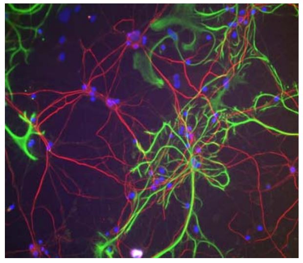
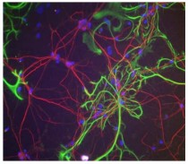
ARG52351 anti-Neurofilament NF-M antibody ICC/IF image
Immunofluorescence: cultured rat neurons and glia stained with ARG52351 anti-Neurofilament NF-M antibody red.


KDF1 Promoted Proliferation, Migration and Invasion of Lung Adenocarcinoma Cells through Activating STAT3 and AKT Pathway
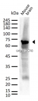
ARG65350: WB /

 New Products