


MyoD1 Rabbit Monoclonal Antibody (ARB860)
 Datasheet

Key features and details
- Target:MyoD1
- Host Species:rabbit monoclonal antibody
- Molecular Weight:35 kDa
- Purity:ProA affinity purified IgG
- Species Cross - reactivity:Human
- Applications:IHC - P
- Swissprot ID:P15172
-
Brand:

CAT.NO. : ARB6652
US$ Please choose
US$ Please choose
Size:
Trail, Bulk size or Custom requests Please contact us
*产品价格可能会有所调整,请以品牌方官网实时更新的价格为准,以确保准确性。
 Product Details
Background
MyoD1, one of the MyoD family of myogenic helix - loop - helix transcription factors, combined with myogenin, plays a role in coordinating the myogenic differentiation pathway from the determination of mesodermal precursors into myoblasts, the differentiation of myoblasts into myotubes, and finally the maturation of myotubes into skeletal myofibers. Normal mature skeletal muscle does not express MyoD1 protein. MyoD1 is expressed in myoblasts before differentiation while myogenin has post - differentiation functions. Anti - MyoD1 immunostaining identifies cells committed to myogenesis in their earliest phase, thus, it is a better biomarker for less differentiated Rhabdomyosarcoma cells (RMS).
RMS are the most frequent malignant soft tissue neoplasms of childhood. While better differentiated RMS have cross - striations or rhabdomyoblasts that allow for a confident morphologic diagnosis, less differentiated RMS resemble other small blue round - cell tumors.
Studies suggest, anti - MyoD1 may be used together with anti - myogenin and anti - desmin as a panel of markers since any RMS is virtually never negative for all three markers simultaneously.
Overview
| Target | MyoD1 |
| Host Species | rabbit monoclonal antibody |
| Molecular Weight | 35 kDa |
| Purity | ProA affinity purified IgG |
| Species Cross-reactivity | Human |
| Form | Liquid |
| Applications | IHC-P |
| Swissprot ID | P15172 |
| Immunogen | Synthetic peptide corresponding to residues within aa1-100 of Human MyoD1 |
| Storage Buffer | PBS 59%, Sodium azide 0.01%, Glycerol 40%, BSA 0.05% |
| Storage Conditions | -25°C to -18°C |
| Dilutions | IHC-P: 1:100-1:200 |
| Subcellular Location | Nucleus |
| Recommended Method | Heat induced epitope retrieval with Tris-EDTA buffer (pH 9.0), primary antibody incubate at RT (18°C-25°C) for 30 minutes |
Data
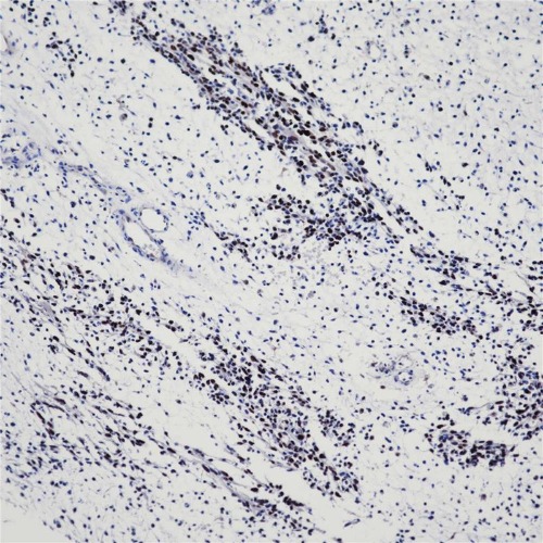
Immunohistochemistry (Formalin/PFA - fixed, paraffin - embedded sections) analysis of Rhabdomyosarcoma tissue labeling Myo D1 with ARA860. Heat mediated antigen retrieval was performed using Tris/EDTA buffer pH 9.0.
Storage
Store at -20°C. Stable for one year from the date of shipment.
Research Use Only
For Research Use Only. Not for use in diagnostic procedures.
New Products


