


Senescence Marker Antibody Panel


Key features and details
- 产品描述:
- 靶点名称:
-
Brand:

 Product Details
| 货号 | 内含物名称 | 宿主克隆性 | 反应 | 应用 | 包装 |
|---|---|---|---|---|---|
| ARG42668 | anti-CDKN2A / p16INK4a antibody | Rabbit pAb | Hu, Ms, Rat | ICC/IF, IP, WB | 20 μl |
| ARG51632 | anti-Rb1 / Retinoblastoma protein phospho (Ser807) antibody | Rabbit pAb | Hu, Ms, Rat | IHC-P, WB | 20 μl |
| ARG10519 | anti-p53 antibody [Pab1801] | Mouse mAb | Hu, Ms, Rat | ChIP, ELISA, FACS, ICC/IF, IHC-P, IHC-Fr, IP, RIA, WB | 20 μg |
| ARG57928 | anti-p21 antibody | Rabbit pAb | Hu, Ms, Rat | ICC/IF, WB | 20 μl |
| 产品描述 | Senescence Marker Antibody Panel is an all-in-one solution to make senescence research easy and economic. The hallmark of senescence is cell cycle arrest. This antibody panel comprises antibodies for detecting senescence-associated cell cycle arrest including p16 INK4a-Rb and p53-p21 pathways. It is the best solution to study senescence and cell cycle arrest in human, mouse, and rat samples. Related news: Senescence Marker Antibody Panel is launched |
|---|---|
| 靶点名称 | Senescence Marker |
| 存放说明 | For continuous use, store undiluted antibody at 2-8°C for up to a week. For long-term storage, aliquot and store at -20°C or below. Storage in frost free freezers is not recommended. Avoid repeated freeze/thaw cycles. Suggest spin the vial prior to opening. The antibody solution should be gently mixed before use. |
|---|---|
| 注意事项 | For laboratory research only, not for drug, diagnostic or other use. |
| 产品亮点 | Related products: Senescence antibodies; Senescence ELISA Kits; Senescence Duos / Panels; |
|---|
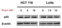
ARG10519 anti-p53 antibody [Pab1801] WB image
Western blot: 20 μg of HCT 116 and LoVo cell lysates stained with ARG10519 anti-p53 antibody [Pab1801] at 1:1000 dilution, overnight at 4°C.

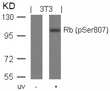
ARG51632 anti-Rb1 / Retinoblastoma protein phospho (Ser807) antibody WB image
Western blot: Extracts from 3T3 cells untreated or treated with UV stained with ARG51632 anti-Rb1 / Retinoblastoma protein phospho (Ser807) antibody.
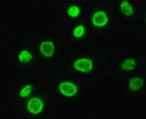
ARG57928 anti-p21 antibody ICC/IF image
Immunofluorescence: HeLa cells stained with ARG57928 anti-p21 antibody at 1:100 dilution.

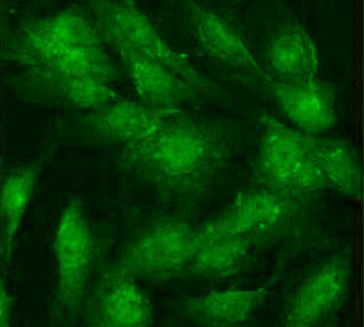
ARG42668 anti-CDKN2A / p16INK4a antibody ICC/IF image
Immunofluorescence: C6 cells stained with ARG42668 anti-CDKN2A / p16INK4a antibody at 1:100 dilution.
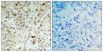
ARG51632 anti-Rb1 / Retinoblastoma protein phospho (Ser807) antibody IHC-P image
Immunohistochemistry: Paraffin-embedded Human breast carcinoma tissue stained with ARG51632 anti-Rb1 / Retinoblastoma protein phospho (Ser807) antibody (left) or the same antibody preincubated with blocking peptide (right).
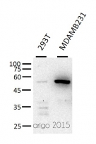
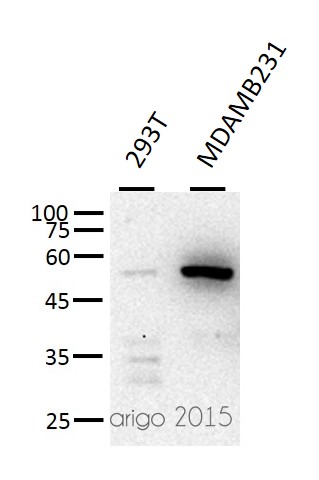
ARG10519 anti-p53 antibody [Pab1801] WB image
Western blot: 30 µg of 293T and MDAMB231 cell lysates stained with ARG10519 anti-p53 antibody [Pab1801] at 1:500 dilution.
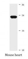
ARG57928 anti-p21 antibody WB image
Western blot: 25 µg of Mouse heart lysate stained with ARG57928 anti-p21 antibody at 1:1000 dilution.
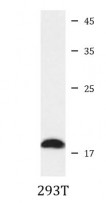
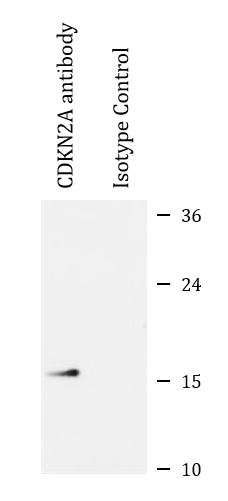
ARG42668 anti-CDKN2A / p16INK4a antibody WB image
Western blot: 25 µg of 293T cell lysate stained with ARG42668 anti-CDKN2A / p16INK4a antibody.
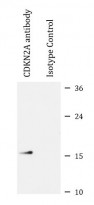
ARG42668 anti-CDKN2A / p16INK4a antibody IP image
Immunoprecipitation: 200 µg extracts of 293T cells were immunoprecipitated and stained with ARG42668 anti-CDKN2A / p16INK4a antibody at 1:1000 dilution.

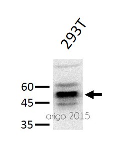
ARG10519 anti-p53 antibody [Pab1801] WB image
Western blot: 20 µg of 293T cell lysate stained with ARG10519 anti-p53 antibody [Pab1801] at 1:500 dilution.
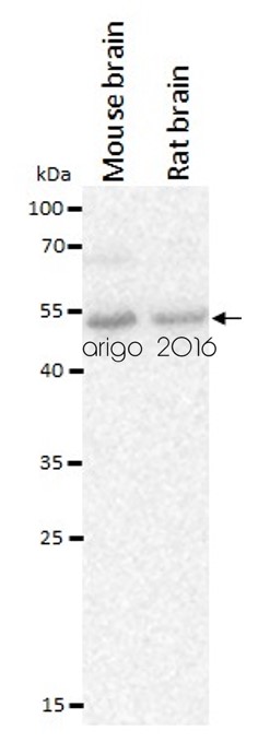
ARG10519 anti-p53 antibody [Pab1801] WB image
Western blot: 20 µg of Mouse brain and Rat brain lysates stained with ARG10519 anti-p53 antibody [Pab1801] at 2 µg/ml dilution.


Human amniotic fluid mesenchymal stem cells attenuate pancreatic cancer cell proliferation and tumor growth in an orthotopic xenograft mouse model
ARG57928: WB / Human

 New Products


