


Aldose Reductase Rabbit Polyclonal Antibody


Key features and details
- Reactivity:Human,Mouse,Rat,Monkey
- Application:WB,IHC,IF/IC
- Host:Rabbit
- Clonality:Polyclonal
- Target:Aldose Reductase
-
Brand:

CAT.NO. : ARA6628
RMB Please choose
RMB Please choose
Size:
Trail, Bulk size or Custom requests Please contact us
*产品价格可能会有所调整,请以品牌方官网实时更新的价格为准,以确保准确性。
 Product Details
Background
Aldose Reductase (also designated ALR2) is member of the monomeric NADPH - dependent aldotetoreductase family. Aldose Reductase catalyzes the reduction of various aldehydes and has been implicated in the development of diabetic complications by catalyzing the reduction of the aldehyde form of glucose, to the corresponding sugar alcohol, sorbitol. This pathway plays a minor role in glucose metabolism in most tissues. However in diabetic hyperglycemia, cells undergoing Insulin - independent uptake of glucose accumulate significant quantities of sorbitol. The resulting hyperosmotic stress to cells may be a cause of diabetic complications such as neuropathy, retinopathy, and cataracts. Aldose Reductase is very similar to human aldehyde reductase (designated ALR1), bovine prostaglandin F synthase and to the European common frog protein, ρ - crystallin.
Application
|
Application |
Dilution Ratio |
|
WB |
1/500 - 1/1000 |
|
IH |
1/100 - 1/200 |
|
IF/IC |
1/100 - 1/500 |
Overview
|
Immunogen |
KLH - conjugated synthetic peptide encompassing a sequence within the C - term region of human Aldose Reductase. The exact sequence is proprietary. |
|
Purification Method |
The antibody was purified by immunogen affinity chromatography. |
|
Clonality |
Polyclonal |
|
Product Form |
Liquid in 0.42% Potassium phosphate, 0.87% Sodium chloride, pH 7.3, 30% glycerol, and 0.01% sodium azide. |
|
Gene Name |
AKR1B1 |
|
Related Names |
ALDR1; Aldose reductase; AR; Aldehyde reductase; Aldo - keto - reductase family 1 member B1 |
|
Gene ID (Human) |
231 |
|
Gene ID (Mouse) |
11677 |
|
Gene ID (Rat) |
24192 |
|
Protein ID (Human) |
P15121 |
|
Protein ID (Mouse) |
P45376 |
|
Protein ID (Rat) |
P07943 |
Data
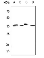
Western blot analysis of Aldose Reductase expression in HEK293T (A), Hela (B), mouse testis (C), rat testis (D) whole cell lysates. (Predicted band size: 35 kD; Observed band size: 36 kD)
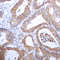
Immunohistochemical analysis of Aldose Reductase staining in human colon cancer formalin fixed paraffin embedded tissue section. The section was pre-treated using heat mediated antigen retrieval with sodium citrate buffer (pH 6.0). The section was then incubated with the antibody at room temperature and detected using an HRP conjugated compact polymer system. DAB was used as the chromogen. The section was then counterstained with haematoxylin and mounted with DPX.
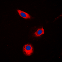
Immunofluorescent analysis of Aldose Reductase staining in Jurkat cells. Formalin-fixed cells were permeabilized with 0.1% Triton X-100 in TBS for 5-10 minutes and blocked with 3% BSA-PBS for 30 minutes at room temperature. Cells were probed with the primary antibody in 3% BSA-PBS and incubated overnight at 4 °C in a humidified chamber. Cells were washed with PBST and incubated with a DyLight 594-conjugated secondary antibody (red) in PBS at room temperature in the dark. DAPI was used to stain the cell nuclei (blue).
Storage
Store at 4°C short term. For long term storage, store at -20°C, avoiding freeze/thaw cycles.
New Products


