


Alpha-1D Adrenergic Receptor Rabbit Polyclonal Antibody

Key features and details
- Reactivity:Human
- Application:WB, IHC, IF/ICC
- Host:Rabbit
- Clonality:Polyclonal
- Target:Alpha-1D Adrenergic Receptor
-
Brand:

 Product Details
|
Application |
Dilution Ratio |
|
WB |
1:500 - 1:2000 |
|
IHC |
1:50 - 1:200 |
|
IF/ICC |
1:50 - 1:100 |
|
Immunogen |
KLH - conjugated synthetic peptide encompassing a sequence within the C - term region of human Alpha - 1D Adrenergic Receptor. The exact sequence is proprietary. |
|
Purification Method |
The antibody was purified by immunogen affinity chromatography. |
|
Clone Type |
Polyclonal |
|
Product Form |
Liquid in 0.42% Potassium phosphate, 0.87% Sodium chloride, pH 7.3, 30% glycerol, and 0.01% sodium azide. |
|
Gene Name |
ADRA1D |
|
Related Names |
ADRA1A; Alpha - 1D adrenergic receptor; Alpha - 1A adrenergic receptor; Alpha - 1D adrenoreceptor; Alpha - 1D adrenoceptor; Alpha - adrenergic receptor 1a |
|
Gene ID (Human) |
146 |
|
Gene ID (Rat) |
29413 |
|
Protein ID (Human) |
P25100 |
|
Protein ID (Mouse) |
P97714 |
|
Protein ID (Rat) |
P23944 |

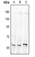
Western blot analysis of Alpha-1D Adrenergic Receptor expression in H9C2 (A), Raw264.7 (B), U87MG (C) whole cell lysates. (Predicted band size: 60 kD; Observed band size: 60 kD)

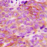
Immunohistochemical analysis of Alpha-1D Adrenergic Receptor staining in human breast cancer formalin fixed paraffin embedded tissue section. The section was pre-treated using heat mediated antigen retrieval with sodium citrate buffer (pH 6.0). The section was then incubated with the antibody at room temperature and detected using an HRP conjugated compact polymer system. DAB was used as the chromogen. The section was then counterstained with haematoxylin and mounted with DPX.
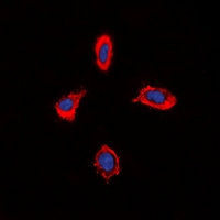
Immunofluorescent analysis of Alpha-1D Adrenergic Receptor staining in MCF7 cells. Formalin-fixed cells were permeabilized with 0.1% Triton X-100 in TBS for 5-10 minutes and blocked with 3% BSA-PBS for 30 minutes at room temperature. Cells were probed with the primary antibody in 3% BSA-PBS and incubated overnight at 4 °C in a hidified chamber. Cells were washed with PBST and incubated with a DyLight 594-conjugated secondary antibody (red) in PBS at room temperature in the dark. DAPI was used to stain the cell nuclei (blue).
New Products


