


anti-ADO antibody


Key features and details
- 产品描述:
- 反应物种:
- 应用:
- 宿主:
- 克隆:
- 同位型:
- 靶点名称:
- 抗原物种:
- 抗原:
-
Brand:

 Product Details
| 产品描述 | Rabbit Polyclonal antibody recognizes ADO |
|---|---|
| 反应物种 | Hu, Ms, Rat |
| 应用 | FACS, ICC/IF, IHC-P, WB |
| 宿主 | Rabbit |
| 克隆 | Polyclonal |
| 同位型 | IgG |
| 靶点名称 | ADO |
| 抗原物种 | Human |
| 抗原 | Recombinant protein corresponding to E49-E261 of Human ADO. |
| 偶联标记 | Un-conjugated |
| 別名 | 2-aminoethanethiol dioxygenase; C10orf22; EC 1.13.11.19; Cysteamine dioxygenase |
| 应用建议 |
| ||||||||||
|---|---|---|---|---|---|---|---|---|---|---|---|
| 应用说明 | IHC-P: Antigen Retrieval: Heat mediation was performed in EDTA buffer (pH 8.0). * The dilutions indicate recommended starting dilutions and the optimal dilutions or concentrations should be determined by the scientist. |
| 形式 | Liquid |
|---|---|
| 纯化 | Affinity purification with immunogen. |
| 缓冲液 | 0.9% NaCl, 0.2% Na2HPO4, 0.05% Sodium azide and 5% BSA. |
| 抗菌剂 | 0.05% Sodium azide |
| 稳定剂 | 5% BSA |
| 浓度 | 0.5 mg/ml |
| 存放说明 | For continuous use, store undiluted antibody at 2-8°C for up to a week. For long-term storage, aliquot and store at -20°C or below. Storage in frost free freezers is not recommended. Avoid repeated freeze/thaw cycles. Suggest spin the vial prior to opening. The antibody solution should be gently mixed before use. |
| 注意事项 | For laboratory research only, not for drug, diagnostic or other use. |
| 数据库连接 | |
|---|---|
| 基因名称 | ADO |
| 全名 | 2-aminoethanethiol (cysteamine) dioxygenase |
| 背景介绍 | Human thiol dioxygenases include cysteine dioxygenase (CDO; MIM 603943) and cysteamine (2-aminoethanethiol) dioxygenase (ADO; EC 1.13.11.19). CDO adds 2 oxygen atoms to free cysteine, whereas ADO adds 2 oxygen atoms to free cysteamine to form hypotaurine (Dominy et al., 2007 [PubMed 17581819]).[supplied by OMIM, Mar 2008] |
| 预测分子量 | 30 kDa |

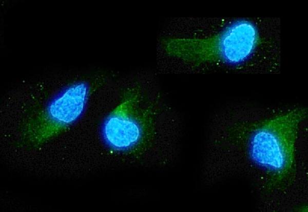
ARG59452 anti-ADO antibody ICC/IF image
Immunofluorescence: A549 cells were blocked with 10% goat serum and then stained with ARG59452 anti-ADO antibody (green) at 2 µg/ml dilution, overnight at 4°C. DAPI (blue) for nuclear staining.
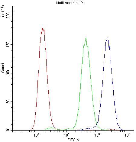
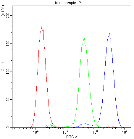
ARG59452 anti-ADO antibody FACS image
Flow Cytometry: A549 cells were blocked with 10% normal goat serum and then stained with ARG59452 anti-ADO antibody (blue) at 1 µg/10^6 cells for 30 min at 20°C, followed by DyLight®488 labelled secondary antibody. Isotype control antibody (green) was rabbit IgG (1 µg/10^6 cells) used under the same conditions. Unlabelled sample (red) was also used as a control.
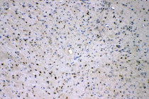
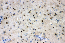
ARG59452 anti-ADO antibody IHC-P image
Immunohistochemistry: Paraffin-embedded Rat brain tissue. Antigen Retrieval: Heat mediation was performed in EDTA buffer (pH 8.0). The tissue section was blocked with 10% goat serum. The tissue section was then stained with ARG59452 anti-ADO antibody at 2 µg/ml dilution, overnight at 4°C.

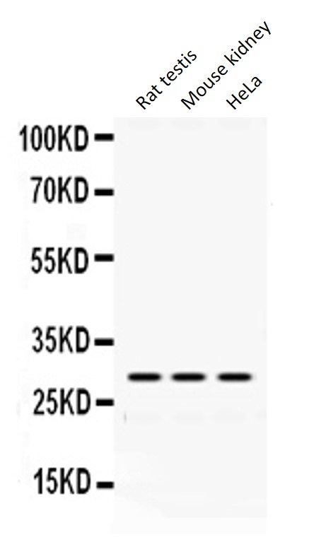
ARG59452 anti-ADO antibody WB image
Western blot: Rat testis, Mouse kidney and HeLa whole cell lysates stained with ARG59452 anti-ADO antibody at 0.5 µg/ml dilution.
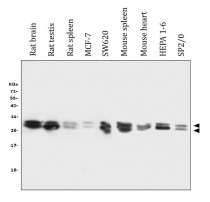
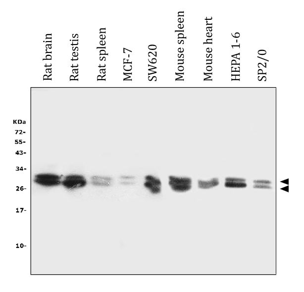
ARG59452 anti-ADO antibody WB image
Western blot: 50 µg of sample under reducing conditions. Rat brain, Rat testis, Rat spleen, MCF-7, SW620, Mouse spleen, Mouse heart, HEPA1-6 and SP2/0 whole cell lysates stained with ARG59452 anti-ADO antibody at 0.5 µg/ml dilution, overnight at 4°C.
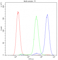
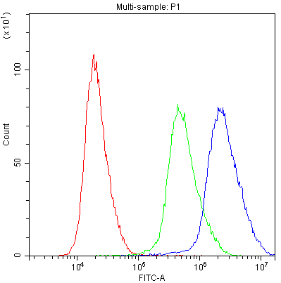
ARG59452 anti-ADO antibody FACS image
Flow Cytometry: U251 cells were blocked with 10% normal goat serum and then stained with ARG59452 anti-ADO antibody (blue) at 1 µg/10^6 cells for 30 min at 20°C, followed by DyLight®488 labelled secondary antibody. Isotype control antibody (green) was rabbit IgG (1 µg/10^6 cells) used under the same conditions. Unlabelled sample (red) was also used as a control.
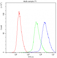
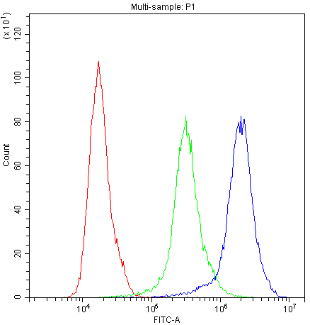
ARG59452 anti-ADO antibody FACS image
Flow Cytometry: HeLa cells were blocked with 10% normal goat serum and then stained with ARG59452 anti-ADO antibody (blue) at 1 µg/10^6 cells for 30 min at 20°C, followed by DyLight®488 labelled secondary antibody. Isotype control antibody (green) was rabbit IgG (1 µg/10^6 cells) used under the same conditions. Unlabelled sample (red) was also used as a control.
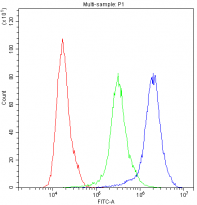
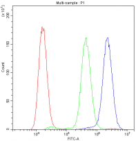
ARG59452 anti-ADO antibody FACS image
Flow Cytometry: PC-3 cells were blocked with 10% normal goat serum and then stained with ARG59452 anti-ADO antibody (blue) at 1 µg/10^6 cells for 30 min at 20°C, followed by DyLight®488 labelled secondary antibody. Isotype control antibody (green) was rabbit IgG (1 µg/10^6 cells) used under the same conditions. Unlabelled sample (red) was also used as a control.
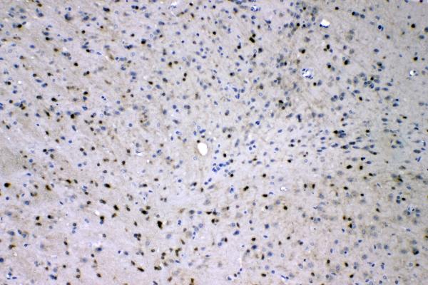
ARG59452 anti-ADO antibody IHC-P image
Immunohistochemistry: Paraffin-embedded Mouse brain tissue. Antigen Retrieval: Heat mediation was performed in EDTA buffer (pH 8.0). The tissue section was blocked with 10% goat serum. The tissue section was then stained with ARG59452 anti-ADO antibody at 2 µg/ml dilution, overnight at 4°C.


 New Products


