


anti-AMFR antibody


Key features and details
- 产品描述:
- 反应物种:
- 应用:
- 宿主:
- 克隆:
- 同位型:
- 靶点名称:
- 抗原物种:
- 抗原:
-
Brand:

 Product Details
| 产品描述 | Rabbit Polyclonal antibody recognizes AMFR |
|---|---|
| 反应物种 | Hu, Ms, Rat |
| 应用 | FACS, IHC-P, WB |
| 宿主 | Rabbit |
| 克隆 | Polyclonal |
| 同位型 | IgG |
| 靶点名称 | AMFR |
| 抗原物种 | Human |
| 抗原 | E. coli-derived Human AMFR recombinant protein (Position: E553-S643). Human AMFR shares 89% amino acid (aa) sequence identity with Mouse AMFR. |
| 偶联标记 | Un-conjugated |
| 別名 | EC 6.3.2.-; E3 ubiquitin-protein ligase AMFR; RNF45; GP78; Autocrine motility factor receptor; gp78; AMF receptor; RING finger protein 45 |
| 应用建议 |
| ||||||||
|---|---|---|---|---|---|---|---|---|---|
| 应用说明 | IHC-P: Antigen Retrieval: Heat mediation was performed in Citrate buffer (pH 6.0) for 20 min. * The dilutions indicate recommended starting dilutions and the optimal dilutions or concentrations should be determined by the scientist. |
| 形式 | Liquid |
|---|---|
| 纯化 | Affinity purification with immunogen. |
| 缓冲液 | 0.9% NaCl, 0.2% Na2HPO4, 0.05% Sodium azide and 5% BSA. |
| 抗菌剂 | 0.05% Sodium azide |
| 稳定剂 | 5% BSA |
| 浓度 | 0.5 mg/ml |
| 存放说明 | For continuous use, store undiluted antibody at 2-8°C for up to a week. For long-term storage, aliquot and store at -20°C or below. Storage in frost free freezers is not recommended. Avoid repeated freeze/thaw cycles. Suggest spin the vial prior to opening. The antibody solution should be gently mixed before use. |
| 注意事项 | For laboratory research only, not for drug, diagnostic or other use. |
| 数据库连接 | |
|---|---|
| 基因名称 | AMFR |
| 全名 | autocrine motility factor receptor, E3 ubiquitin protein ligase |
| 背景介绍 | This locus encodes a glycosylated transmembrane receptor. Its ligand, autocrine motility factor, is a tumor motility-stimulating protein secreted by tumor cells. The encoded receptor is also a member of the E3 ubiquitin ligase family of proteins. It catalyzes ubiquitination and endoplasmic reticulum-associated degradation of specific proteins. [provided by RefSeq, Feb 2012] |
| 生物功能 | E3 ubiquitin-protein ligase that mediates the polyubiquitination of a number of proteins such as CD3D, CYP3A4, CFTR and APOB for proteasomal degradation. Component of a VCP/p97-AMFR/gp78 complex that participates in the final step of endoplasmic reticulum-associated degradation (ERAD). The VCP/p97-AMFR/gp78 complex is involved in the sterol-accelerated ERAD degradation of HMGCR through binding to the HMGCR-INSIG complex at the ER membrane and initiating ubiquitination of HMGCR. The ubiquitinated HMGCR is then released from the ER by the complex into the cytosol for subsequent destruction. Also acts as a scaffold protein to assemble a complex that couples ubiquitination, retranslocation and deglycosylation. Mediates tumor invasion and metastasis as a receptor for the GPI/autocrine motility factor. [UniProt] |
| 细胞定位 | Endoplasmic reticulum membrane; Multi-pass membrane protein. [UniProt] |
| 预测分子量 | 73 kDa |
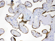
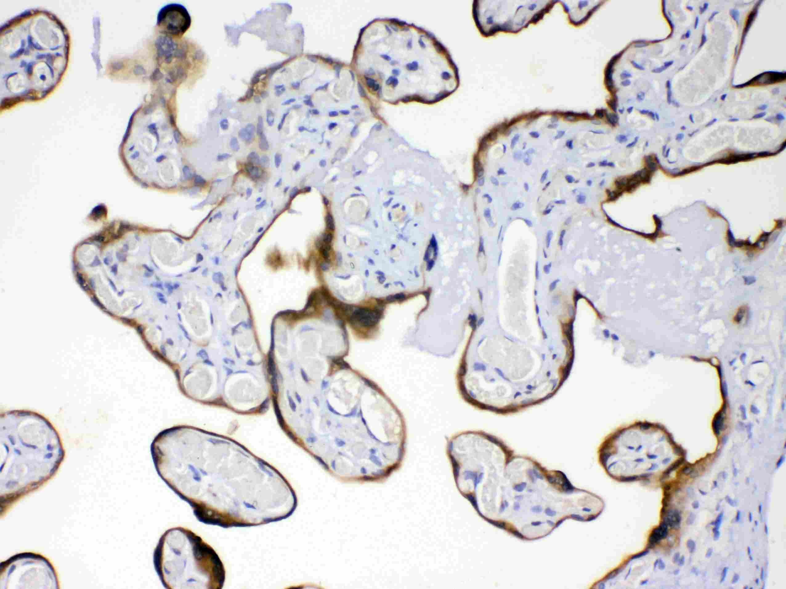
ARG58219 anti-AMFR antibody IHC-P image
Immunohistochemistry: Paraffin-embedded Human placentas stained with ARG58219 anti-AMFR antibody at 1 µg/ml dilution.
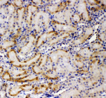
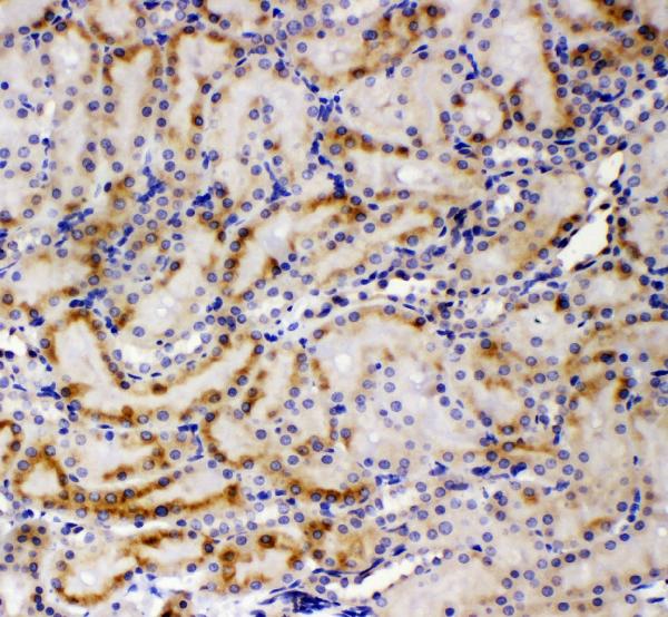
ARG58219 anti-AMFR antibody IHC-P image
Immunohistochemistry: Paraffin-embedded Rat kidney tissue. Antigen Retrieval: Heat mediation was performed in Citrate buffer (pH 6.0) for 20 min. The tissue section was blocked with 10% goat serum. The tissue section was then stained with ARG58219 anti-AMFR antibody at 1 µg/ml dilution, overnight at 4°C.
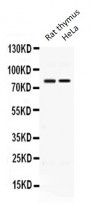
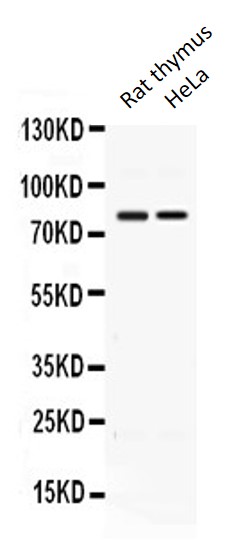
ARG58219 anti-AMFR antibody WB image
Western blot: Rat thymus extract and HeLa whole cell lysate stained with ARG58219 anti-AMFR antibody at 0.5 µg/ml dilution.
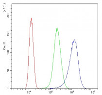
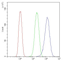
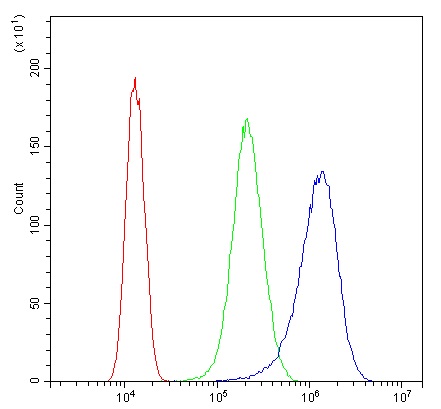
ARG58219 anti-AMFR antibody FACS image
Flow Cytometry: SiHa cells were blocked with 10% normal goat serum and then stained with ARG58219 anti-AMFR antibody (blue) at 1 µg/10^6 cells for 30 min at 20°C, followed by incubation with DyLight®488 labelled secondary antibody. Isotype control antibody (green) was rabbit IgG (1 µg/10^6 cells) used under the same conditions. Unlabelled sample (red) was also used as a control.
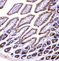
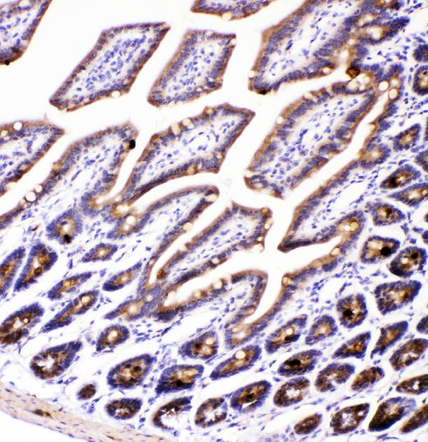
ARG58219 anti-AMFR antibody IHC-P image
Immunohistochemistry: Paraffin-embedded Mouse intestine tissue. Antigen Retrieval: Heat mediation was performed in Citrate buffer (pH 6.0) for 20 min. The tissue section was blocked with 10% goat serum. The tissue section was then stained with ARG58219 anti-AMFR antibody at 1 µg/ml dilution, overnight at 4°C.
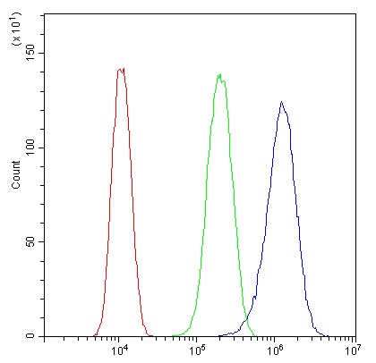
ARG58219 anti-AMFR antibody FACS image
Flow Cytometry: U87 cells were blocked with 10% normal goat serum and then stained with ARG58219 anti-AMFR antibody (blue) at 1 µg/10^6 cells for 30 min at 20°C, followed by incubation with DyLight®488 labelled secondary antibody. Isotype control antibody (green) was rabbit IgG (1 µg/10^6 cells) used under the same conditions. Unlabelled sample (red) was also used as a control.


 New Products


