


anti-ATP5G1 + ATP5G2 + ATP5G3 antibody


Key features and details
- 产品描述:
- 反应物种:
- 应用:
- 宿主:
- 克隆:
- 同位型:
- 靶点名称:
- 抗原物种:
- 抗原:
-
Brand:

 Product Details
| 产品描述 | Rabbit Polyclonal antibody recognizes ATP5G1 + ATP5G2 + ATP5G3 |
|---|---|
| 反应物种 | Hu, Ms, Rat |
| 应用 | FACS, IHC-P, WB |
| 宿主 | Rabbit |
| 克隆 | Polyclonal |
| 同位型 | IgG |
| 靶点名称 | ATP5G1 + ATP5G2 + ATP5G3 |
| 抗原物种 | Human |
| 抗原 | Recombinant protein corresponding to D62-L113 of Human ATP5G1. |
| 偶联标记 | Un-conjugated |
| 別名 | ATP5G1: ATP5A; ATP5G; ATP synthase F(0) complex subunit C1, mitochondrial; ATP synthase lipid-binding protein; ATP synthase proteolipid P1; ATP synthase proton-transporting mitochondrial F(0) complex subunit C1; ATPase protein 9; ATPase subunit c ATP5G2: ATP5A; ATP synthase F(0) complex subunit C2, mitochondrial; ATP synthase lipid-binding protein; ATP synthase proteolipid P2; ATP synthase proton-transporting mitochondrial F(0) complex subunit C2; ATPase protein 9; ATPase subunit c ATP5G3: P3; ATP synthase F(0) complex subunit C3, mitochondrial; ATP synthase lipid-binding protein; ATP synthase proteolipid P3; ATP synthase proton-transporting mitochondrial F(0) complex subunit C3; ATPase protein 9; ATPase subunit c |
| 应用建议 |
| ||||||||
|---|---|---|---|---|---|---|---|---|---|
| 应用说明 | * The dilutions indicate recommended starting dilutions and the optimal dilutions or concentrations should be determined by the scientist. |
| 形式 | Liquid |
|---|---|
| 纯化 | Affinity purification with immunogen. |
| 缓冲液 | 0.2% Na2HPO4, 0.9% NaCl, 0.05% Sodium azide and 4% Trehalose. |
| 抗菌剂 | 0.05% Sodium azide |
| 稳定剂 | 4% Trehalose |
| 浓度 | 0.5 mg/ml |
| 存放说明 | For continuous use, store undiluted antibody at 2-8°C for up to a week. For long-term storage, aliquot and store at -20°C or below. Storage in frost free freezers is not recommended. Avoid repeated freeze/thaw cycles. Suggest spin the vial prior to opening. The antibody solution should be gently mixed before use. |
| 注意事项 | For laboratory research only, not for drug, diagnostic or other use. |
| 基因名称 | ATP5G1; ATP5G2; ATP5G3 |
|---|---|
| 全名 | ATP synthase, H+ transporting, mitochondrial Fo complex, subunit C1 (subunit 9) ATP synthase, H+ transporting, mitochondrial Fo complex, subunit C2 (subunit 9) ATP synthase, H+ transporting, mitochondrial Fo complex, subunit C3 (subunit 9) |
| 背景介绍 | This gene encodes a subunit of mitochondrial ATP synthase. Mitochondrial ATP synthase catalyzes ATP synthesis, utilizing an electrochemical gradient of protons across the inner membrane during oxidative phosphorylation. ATP synthase is composed of two linked multi-subunit complexes: the soluble catalytic core, F1, and the membrane-spanning component, Fo, comprising the proton channel. The catalytic portion of mitochondrial ATP synthase consists of 5 different subunits (alpha, beta, gamma, delta, and epsilon) assembled with a stoichiometry of 3 alpha, 3 beta, and a single representative of the other 3. The proton channel seems to have nine subunits (a, b, c, d, e, f, g, F6 and 8). This gene is one of three genes that encode subunit c of the proton channel. Each of the three genes have distinct mitochondrial import sequences but encode the identical mature protein. Alternatively spliced transcript variants encoding the same protein have been identified. [provided by RefSeq, Jul 2008] |
| 生物功能 | Mitochondrial membrane ATP synthase (F(1)F(0) ATP synthase or Complex V) produces ATP from ADP in the presence of a proton gradient across the membrane which is generated by electron transport complexes of the respiratory chain. F-type ATPases consist of two structural domains, F(1) - containing the extramembraneous catalytic core and F(0) - containing the membrane proton channel, linked together by a central stalk and a peripheral stalk. During catalysis, ATP synthesis in the catalytic domain of F(1) is coupled via a rotary mechanism of the central stalk subunits to proton translocation. Part of the complex F(0) domain. A homomeric c-ring of probably 10 subunits is part of the complex rotary element. [UniProt] |
| 细胞定位 | ATP5G1/G2/G3: Mitochondrion membrane; Multi-pass membrane protein. [UniProt] |
| 预测分子量 | ATP5G1: 14 kDa ATP5G2: 15 kDa ATP5G3: 15 kDa |
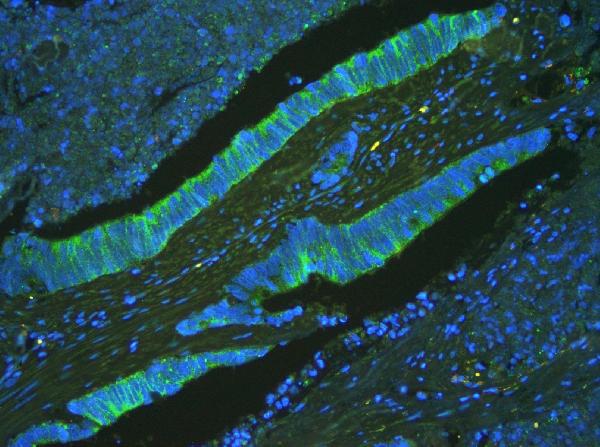
ARG42697 anti-ATP5G1 + ATP5G2 + ATP5G3 antibody IHC-P image
Immunohistochemistry: Paraffin-embedded Human intestinal tissue. Antigen Retrieval: Heat mediation was performed in EDTA buffer (pH 8.0). The tissue section was blocked with 10% goat serum. The tissue section was then stained with ARG42697 anti-ATP5G1 + ATP5G2 + ATP5G3 antibody at 2 µg/ml dilution, overnight at 4°C.
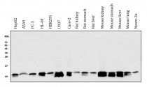
ARG42697 anti-ATP5G1 + ATP5G2 + ATP5G3 antibody WB image
Western blot: 50 µg of sample under reducing conditions. HepG2, A549, PC-3, HL-60, HEK293, U937, Caco-2, Rat kidney, Rat stomach, Rat liver, Mouse kidney, Mouse stomach, Mouse liver, Mouse lung and Neuro-2a whole cell lysates stained with ARG42697 anti-ATP5G1 + ATP5G2 + ATP5G3 antibody at 0.5 µg/ml dilution, overnight at 4°C.

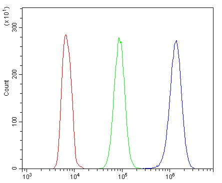
ARG42697 anti-ATP5G1 + ATP5G2 + ATP5G3 antibody FACS image
Flow Cytometry: HL-60 cells were blocked with 10% normal goat serum and then stained with ARG42697 anti-ATP5G1 + ATP5G2 + ATP5G3 antibody (blue) at 1 µg/10^6 cells for 30 min at 20°C, followed by incubation with DyLight®488 labelled secondary antibody. Isotype control antibody (green) was rabbit IgG (1 µg/10^6 cells) used under the same conditions. Unlabelled sample (red) was also used as a control.

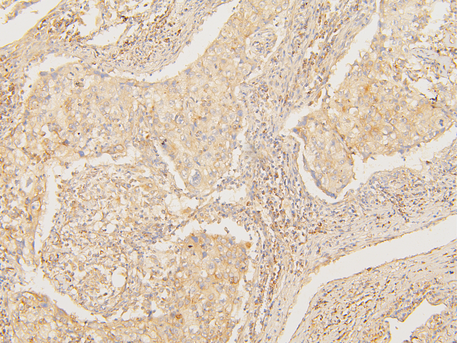
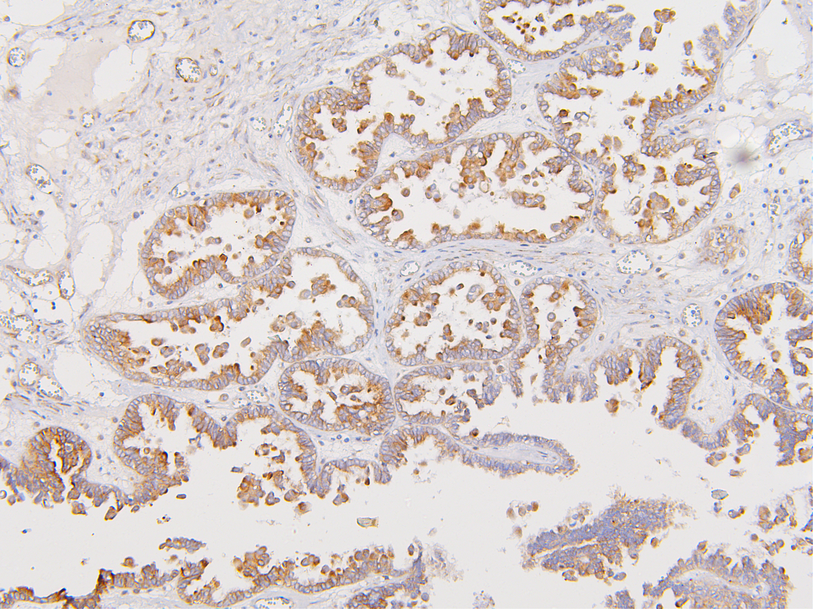
ARG42697 anti-ATP5G1 + ATP5G2 + ATP5G3 antibody IHC-P image
Immunohistochemistry: Paraffin-embedded Human ovarian cancer tissue. Antigen Retrieval: Heat mediation was performed in Citrate buffer (pH 6.0) for 20 min. The tissue section was blocked with 10% goat serum. The tissue section was then stained with ARG42697 anti-ATP5G1 + ATP5G2 + ATP5G3 antibody at 1 µg/ml dilution, overnight at 4°C.
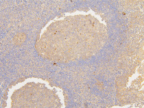
ARG42697 anti-ATP5G1 + ATP5G2 + ATP5G3 antibody IHC-P image
Immunohistochemistry: Paraffin-embedded Human tonsil tissue. Antigen Retrieval: Heat mediation was performed in Citrate buffer (pH 6.0) for 20 min. The tissue section was blocked with 10% goat serum. The tissue section was then stained with ARG42697 anti-ATP5G1 + ATP5G2 + ATP5G3 antibody at 1 µg/ml dilution, overnight at 4°C.
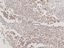
ARG42697 anti-ATP5G1 + ATP5G2 + ATP5G3 antibody IHC-P image
Immunohistochemistry: Paraffin-embedded Human glioma tissue. Antigen Retrieval: Heat mediation was performed in Citrate buffer (pH 6.0) for 20 min. The tissue section was blocked with 10% goat serum. The tissue section was then stained with ARG42697 anti-ATP5G1 + ATP5G2 + ATP5G3 antibody at 1 µg/ml dilution, overnight at 4°C.
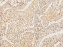
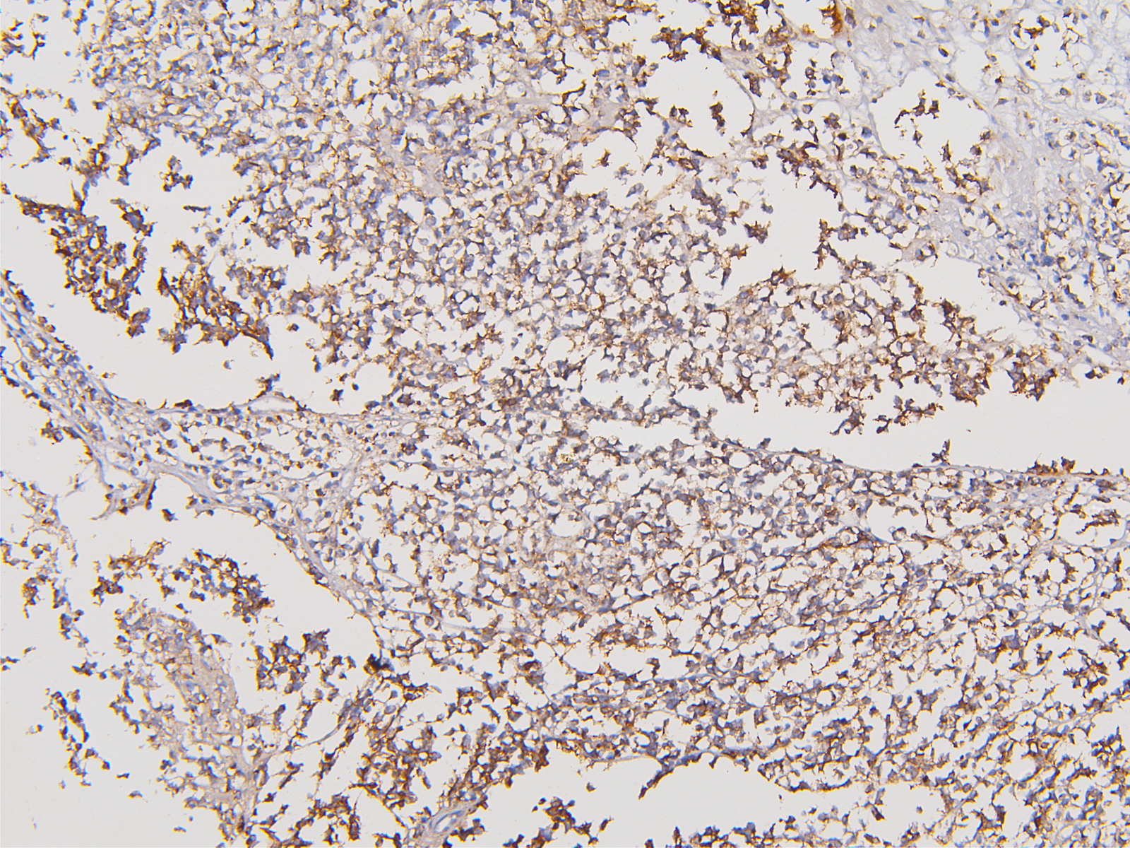
ARG42697 anti-ATP5G1 + ATP5G2 + ATP5G3 antibody IHC-P image
Immunohistochemistry: Paraffin-embedded Human lung cancer tissue. Antigen Retrieval: Heat mediation was performed in Citrate buffer (pH 6.0) for 20 min. The tissue section was blocked with 10% goat serum. The tissue section was then stained with ARG42697 anti-ATP5G1 + ATP5G2 + ATP5G3 antibody at 1 µg/ml dilution, overnight at 4°C.
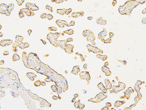
ARG42697 anti-ATP5G1 + ATP5G2 + ATP5G3 antibody IHC-P image
Immunohistochemistry: Paraffin-embedded Human placenta tissue. Antigen Retrieval: Heat mediation was performed in Citrate buffer (pH 6.0) for 20 min. The tissue section was blocked with 10% goat serum. The tissue section was then stained with ARG42697 anti-ATP5G1 + ATP5G2 + ATP5G3 antibody at 1 µg/ml dilution, overnight at 4°C.
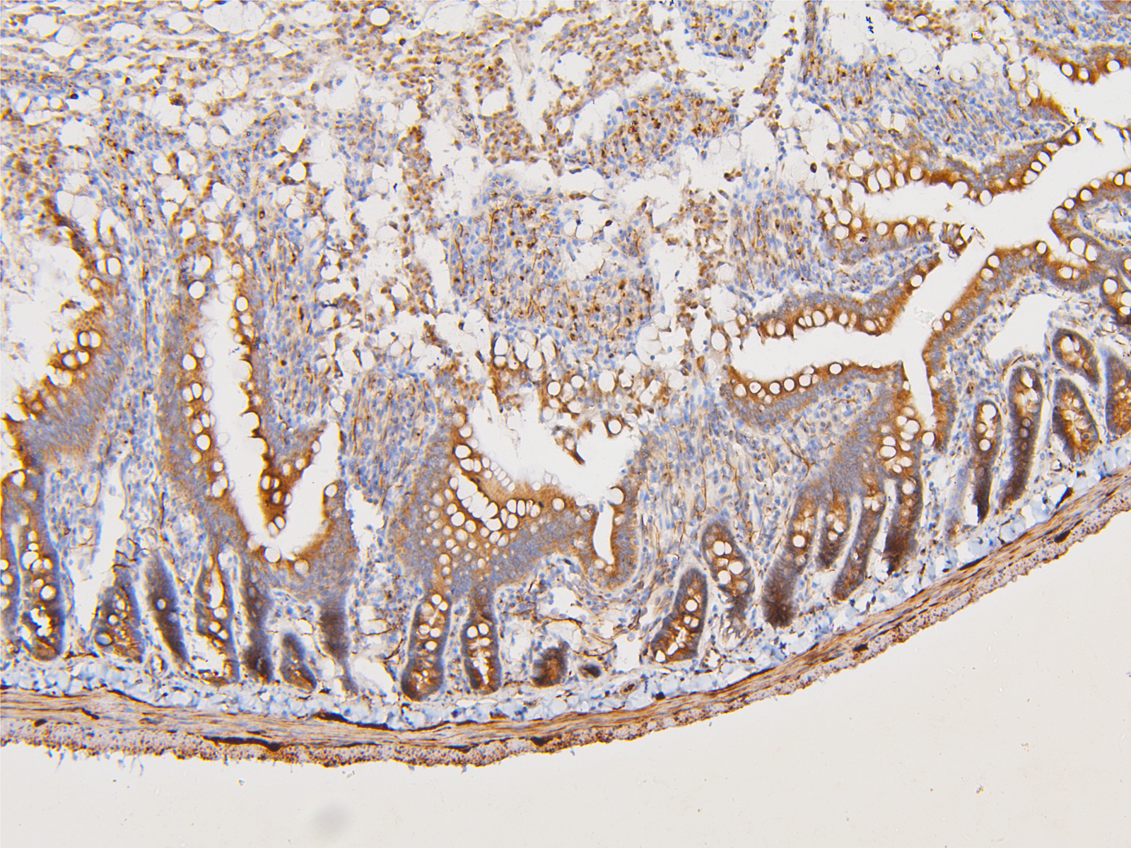
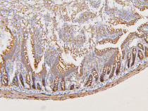
ARG42697 anti-ATP5G1 + ATP5G2 + ATP5G3 antibody IHC-P image
Immunohistochemistry: Paraffin-embedded Mouse intestine tissue. Antigen Retrieval: Heat mediation was performed in Citrate buffer (pH 6.0) for 20 min. The tissue section was blocked with 10% goat serum. The tissue section was then stained with ARG42697 anti-ATP5G1 + ATP5G2 + ATP5G3 antibody at 1 µg/ml dilution, overnight at 4°C.
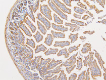
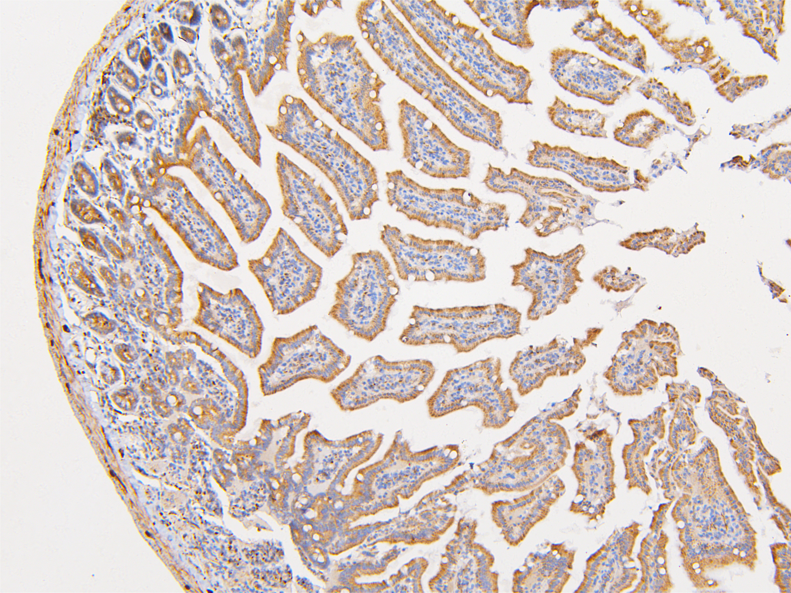
ARG42697 anti-ATP5G1 + ATP5G2 + ATP5G3 antibody IHC-P image
Immunohistochemistry: Paraffin-embedded Rat intestine tissue. Antigen Retrieval: Heat mediation was performed in Citrate buffer (pH 6.0) for 20 min. The tissue section was blocked with 10% goat serum. The tissue section was then stained with ARG42697 anti-ATP5G1 + ATP5G2 + ATP5G3 antibody at 1 µg/ml dilution, overnight at 4°C.
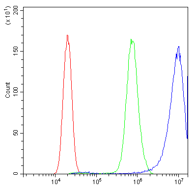
ARG42697 anti-ATP5G1 + ATP5G2 + ATP5G3 antibody FACS image
Flow Cytometry: PC-3 cells were blocked with 10% normal goat serum and then stained with ARG42697 anti-ATP5G1 + ATP5G2 + ATP5G3 antibody (blue) at 1 µg/10^6 cells for 30 min at 20°C, followed by incubation with DyLight®488 labelled secondary antibody. Isotype control antibody (green) was rabbit IgG (1 µg/10^6 cells) used under the same conditions. Unlabelled sample (red) was also used as a control.


 New Products


