


anti-Calnexin antibody


Key features and details
- 产品描述:
- 反应物种:
- 预测物种:
- 应用:
- 特异性:
- 宿主:
- 克隆:
- 同位型:
- 靶点名称:
-
Brand:

 Product Details
| 产品描述 | Goat Polyclonal antibody recognizes Calnexin |
|---|---|
| 反应物种 | Hu, Ms |
| 预测物种 | Cow, Rat, Dog, Pig |
| 应用 | ICC/IF, IHC-P, WB |
| 特异性 | Reported variants represent identical protein (NP_001019820.1, NP_001737.1). |
| 宿主 | Goat |
| 克隆 | Polyclonal |
| 同位型 | IgG |
| 靶点名称 | Calnexin |
| 抗原物种 | Human |
| 抗原 | C-SKTPELNLDQFHDKT |
| 偶联标记 | Un-conjugated |
| 別名 | P90; CNX; p90; Major histocompatibility complex class I antigen-binding protein p88; Calnexin; IP90 |
| 应用建议 |
| ||||||||
|---|---|---|---|---|---|---|---|---|---|
| 应用说明 | WB: Recommend incubate at RT for 1h. IHC-P: Antigen Retrieval: Steam tissue section in Citrate buffer (pH 6.0). * The dilutions indicate recommended starting dilutions and the optimal dilutions or concentrations should be determined by the scientist. | ||||||||
| 阳性对照 | Human cerebellum, Human colorectal cancer, CaCo-2 and NIH/3T3 | ||||||||
| 实际分子量 | 90 - 100 kDa |
| 形式 | Liquid |
|---|---|
| 纯化 | Purified from goat serum by ammonium sulphate precipitation followed by antigen affinity chromatography using the immunizing peptide. |
| 缓冲液 | Tris saline (pH 7.3), 0.02% Sodium azide and 0.5% BSA |
| 抗菌剂 | 0.02% Sodium azide |
| 稳定剂 | 0.5% BSA |
| 浓度 | 0.5 mg/ml |
| 存放说明 | For continuous use, store undiluted antibody at 2-8°C for up to a week. For long-term storage, aliquot and store at -20°C or below. Storage in frost free freezers is not recommended. Avoid repeated freeze/thaw cycles. Suggest spin the vial prior to opening. The antibody solution should be gently mixed before use. |
| 注意事项 | For laboratory research only, not for drug, diagnostic or other use. |
| 数据库连接 | |
|---|---|
| 背景介绍 | This gene encodes a member of the calnexin family of molecular chaperones. The encoded protein is a calcium-binding, endoplasmic reticulum (ER)-associated protein that interacts transiently with newly synthesized N-linked glycoproteins, facilitating protein folding and assembly. It may also play a central role in the quality control of protein folding by retaining incorrectly folded protein subunits within the ER for degradation. Alternatively spliced transcript variants encoding the same protein have been described. [provided by RefSeq, Jul 2008] |
| 研究领域 | Controls and Markers antibody; Neuroscience antibody |
| 预测分子量 | 68 kDa |
| 翻译后修饰 | Phosphorylated at Ser-564 by MAPK3/ERK1. phosphorylation by MAPK3/ERK1 increases its association with ribosomes (By similarity). Palmitoylation by DHHC6 leads to the preferential localization to the perinuclear rough ER. It mediates the association of calnexin with the ribosome-translocon complex (RTC) which is required for efficient folding of glycosylated proteins. Ubiquitinated, leading to proteasomal degradation. Probably ubiquitinated by ZNRF4. |

ARG64980 anti-Calnexin antibody ICC/IF image
Immunofluorescence: Paraformaldehyde fixed NIH/3T3 cells permeabilized with 0.15% Triton. Cells were stained with ARG64980 anti-Calnexin antibody (green) at 10 µg/ml dilution for 1 hour. DAPI (blue) for nuclear staining. Negative control: Unimmunized goat IgG (green) at 10 µg/ml dilution.

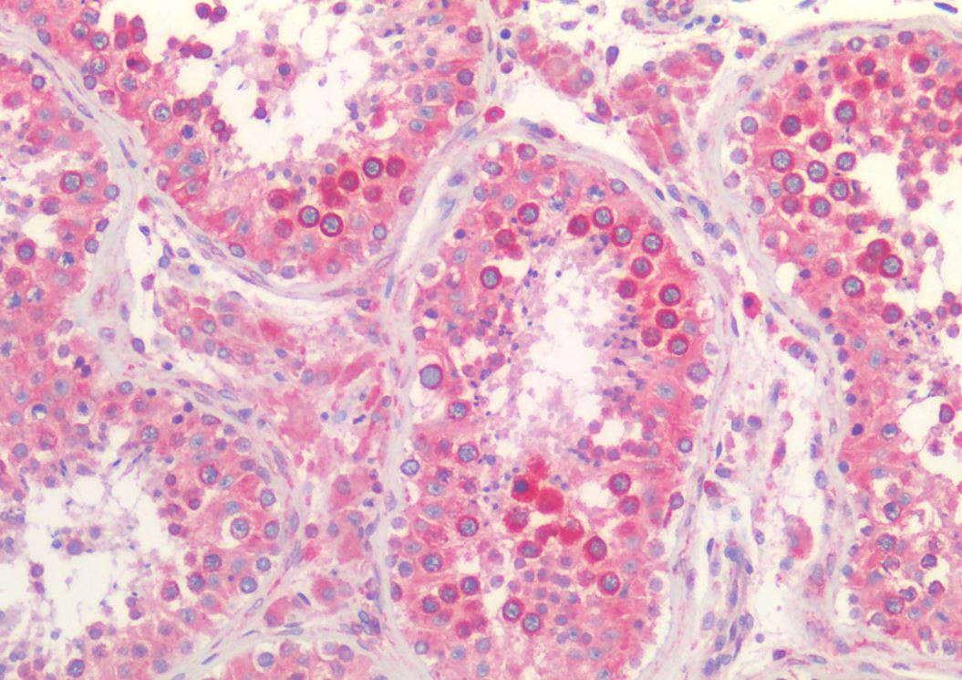
ARG64980 anti-Calnexin antibody IHC-P image
Immunohistochemistry: Paraffin-embedded Human testis tissue. Antigen Retrieval: Steam tissue section in Citrate buffer (pH 6.0). The tissue section was stained with ARG64980 anti-Calnexin antibody at 5 µg/ml dilution followed by AP-staining.

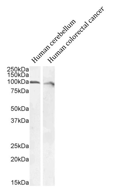
ARG64980 anti-Calnexin antibody WB image
Western blot: 35 µg of Human cerebellum and Human colorectal cancer tissue lysates (in RIPA buffer) stained with ARG64980 anti-Calnexin antibody at 0.1 µg/ml dilution and incubated at RT for 1 hour.
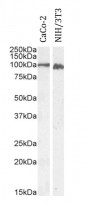
ARG64980 anti-Calnexin antibody WB image
Western blot: 35 µg of CaCo-2 and NIH/3T3 cell lysates (in RIPA buffer) stained with ARG64980 anti-Calnexin antibody at 0.1 and 1 µg/ml dilution, respectively. Both lanes were incubated at RT for 1 hour.

 New Products


