


anti-Calretinin antibody [6A9]
![anti-Calretinin antibody [6A9]](/upload/image/products/ARG10703_IHC_2_210_205.jpg)
![anti-Calretinin antibody [6A9]](/upload/image/products/ARG10703_WB_Rev_RatBrain_170714.jpg)
![anti-Calretinin antibody [6A9]](/upload/image/products/ARG10703_ICC_1.jpg)
![anti-Calretinin antibody [6A9]](/upload/image/products/ARG10703_IHC-Fr_190729.jpg)
![anti-Calretinin antibody [6A9]](/upload/image/products/ARG10703_IHC_1.jpg)
![anti-Calretinin antibody [6A9]](/upload/image/products/ARG10703_WB_171108.jpg)
![anti-Calretinin antibody [6A9]](/upload/image/products/ARG10703_WB_190729.jpg)
![anti-Calretinin antibody [6A9]](/upload/image/products/ARG10703_IHC_2.jpg)
Key features and details
- 产品描述:
- 反应物种:
- 应用:
- 宿主:
- 克隆:
- 克隆号:
- 同位型:
- 靶点名称:
- 抗原物种:
-
Brand:

 Product Details
| 产品描述 | Mouse Monoclonal antibody [6A9] recognizes Calretinin |
|---|---|
| 反应物种 | Hu, Ms, Rat, Cow |
| 应用 | ICC/IF, IHC-Fr, WB |
| 宿主 | Mouse |
| 克隆 | Monoclonal |
| 克隆号 | 6A9 |
| 同位型 | IgA |
| 靶点名称 | Calretinin |
| 抗原物种 | Human |
| 抗原 | Full-length recombinant Human protein. |
| 偶联标记 | Un-conjugated |
| 別名 | CAB29; CR; CAL2; 29 kDa calbindin; Calretinin |
| 应用建议 |
| ||||||||
|---|---|---|---|---|---|---|---|---|---|
| 应用说明 | * The dilutions indicate recommended starting dilutions and the optimal dilutions or concentrations should be determined by the scientist. |
| 形式 | Liquid |
|---|---|
| 纯化 | Affinity purification. |
| 缓冲液 | PBS, 5 mM Sodium azide and 50% Glycerol. |
| 抗菌剂 | 5 mM Sodium azide |
| 稳定剂 | 50% Glycerol |
| 浓度 | 1 mg/ml |
| 存放说明 | For continuous use, store undiluted antibody at 2-8°C for up to a week. For long-term storage, aliquot and store at -20°C. Storage in frost free freezers is not recommended. Avoid repeated freeze/thaw cycles. Suggest spin the vial prior to opening. The antibody solution should be gently mixed before use. |
| 注意事项 | For laboratory research only, not for drug, diagnostic or other use. |
| 数据库连接 | |
|---|---|
| 基因名称 | CALB2 |
| 全名 | calbindin 2 |
| 背景介绍 | This gene encodes an intracellular calcium-binding protein belonging to the troponin C superfamily. Members of this protein family have six EF-hand domains which bind calcium. This protein plays a role in diverse cellular functions, including message targeting and intracellular calcium buffering. It also functions as a modulator of neuronal excitability, and is a diagnostic marker for some human diseases, including Hirschsprung disease and some cancers. Alternative splicing results in multiple transcript variants. [provided by RefSeq, Jun 2010] |
| 生物功能 | Calretinin is a calcium-binding protein which is abundant in auditory neurons. [UniProt] |
| 预测分子量 | 32 kDa |
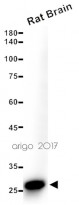
ARG10703 anti-Calretinin antibody [6A9] WB image
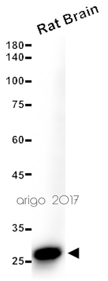
Western blot: 15 µg of Rat brain lysate stained with ARG10703 anti-Calretinin antibody [6A9] at 1:2500 dilution.
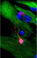
ARG10703 anti-Calretinin antibody [6A9] ICC/IF image
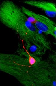
Immunocytochemistry: Rat mixed neuron / glial cultures stained with ARG10703 anti-Calretinin antibody [6A9] (red) at 1:2000, and co-stained with chicken polyclonal antibody to Vimentin (green). Calretinin is prominently expressed in small number of interneurons, while astrocytes and fibroblasts were visualized with the vimentin antibody.
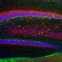
ARG10703 anti-Calretinin antibody [6A9] IHC-Fr image
Immunohistochemistry: Frozen section of Rat hippocampus stained with ARG10703 anti-Calretinin antibody [6A9] (green) at 1:2000 dilution and costained with ARG10686 anti-Parvalbumin antibody (red) at 1:1000 dilution. DAPI (blue) for nuclear staining. (Sample preparation: Following transcardial perfusion of Rat with 4% paraformaldehyde, brain was post fixed for 24 hours, cut to 45 µM, and free-floating sections were stained with above antibodies.)

ARG10703 anti-Calretinin antibody [6A9] IHC-Fr image
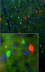
Immunohistochemistry: Frozen sections of adult Rat cortical (45 µM; fixed by transcardial perfusion with 4% paraformaldehyde) was stained with ARG10703 anti-Calretinin antibody [6A9] at 1:1000 (red) and co-stained with chicken polyclonal antibody to calbinidin (green). In the motor cortex, calretinin is expressed in a small population of interneurons that do not express calbindin. Because each antibody specifically labels a different population of cells exclusively, the cells are either stained with red, or green.
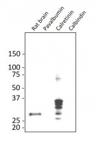
ARG10703 anti-Calretinin antibody [6A9] WB image
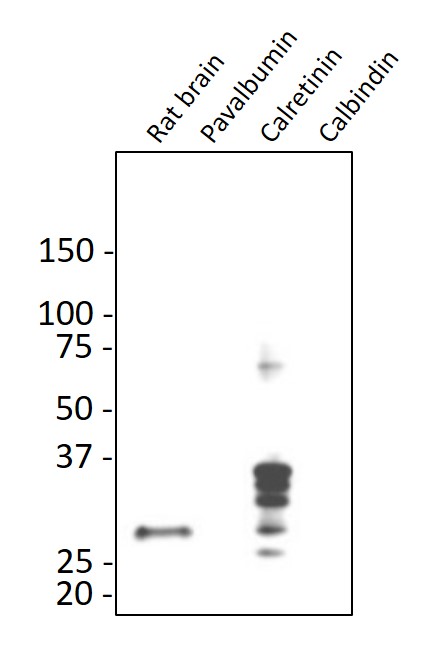
Western blot: Rat brain, pavalbumin, calretinin, and calbindin recombinant proteins was stained with ARG10703 anti-Calretinin antibody [6A9] at 1:5000 dilution.
ARG10703 anti-Calretinin antibody [6A9] WB image
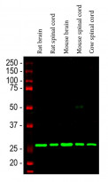
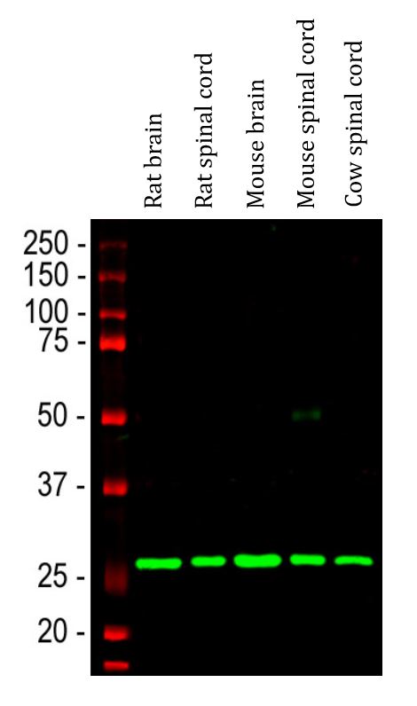
Western blot: Rat brain, Rat spinal cord, Mouse brain, Mouse spinal cord and Cow spinal cord lysates stained with ARG10703 anti-Calretinin antibody [6A9] (green) at 1:2000 dilution.
ARG10703 anti-Calretinin antibody [6A9] IHC-Fr image
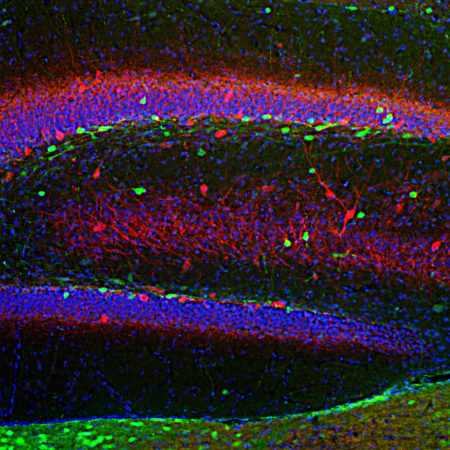
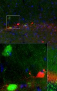
Immunohistochemistry: Frozen sections of adult Mouse brain hippocampal (45 µM; fixed by transcardial perfusion with 4% paraformaldehyde) was stained with ARG10703 anti-Calretinin antibody [6A9] at 1:1000 (red) and co-stained with antibody to calbinidin (green). In the stratum radiatum of CA1 region, calretinin expresses in a small number of interneurons that do not express calbindin. As a result, our antibodies label different neurons in either red or green. Insets are high-magnification images of the boxed area in each picture. Blue is a hoechst DNA staining.
 New Products


