


anti-CD209 / DC-SIGN antibody

Key features and details
- 产品描述:
- 反应物种:
- 应用:
- 宿主:
- 克隆:
- 同位型:
- 靶点名称:
- 抗原物种:
- 抗原:
-
Brand:

 Product Details
| 产品描述 | Rabbit Polyclonal antibody recognizes CD209 / DC-SIGN |
|---|---|
| 反应物种 | Hu |
| 应用 | FACS, IHC-P, WB |
| 宿主 | Rabbit |
| 克隆 | Polyclonal |
| 同位型 | IgG |
| 靶点名称 | CD209 / DC-SIGN |
| 抗原物种 | Human |
| 抗原 | Synthetic peptide corresponding to a sequence of Human CD209 / DC-SIGN. (MSDSKEPRLQQLGLLEEEQLRGLGFRQTRGYKSLA) |
| 偶联标记 | Un-conjugated |
| 別名 | CDSIGN; Dendritic cell-specific ICAM-3-grabbing non-integrin 1; CLEC4L; DC-SIGN; CD antigen CD209; CD209 antigen; DC-SIGN1; C-type lectin domain family 4 member L |
| 应用建议 |
| ||||||||
|---|---|---|---|---|---|---|---|---|---|
| 应用说明 | IHC-P: Antigen Retrieval: Heat mediation was performed in Citrate buffer (pH 6.0) for 20 min. * The dilutions indicate recommended starting dilutions and the optimal dilutions or concentrations should be determined by the scientist. | ||||||||
| 实际分子量 | ~ 46 kDa |
| 形式 | Liquid |
|---|---|
| 纯化 | Affinity purification with immunogen. |
| 缓冲液 | 0.2% Na2HPO4, 0.9% NaCl, 0.05% Sodium azide and 4% Trehalose. |
| 抗菌剂 | 0.05% Sodium azide |
| 稳定剂 | 4% Trehalose |
| 浓度 | 0.5 mg/ml |
| 存放说明 | For continuous use, store undiluted antibody at 2-8°C for up to a week. For long-term storage, aliquot and store at -20°C or below. Storage in frost free freezers is not recommended. Avoid repeated freeze/thaw cycles. Suggest spin the vial prior to opening. The antibody solution should be gently mixed before use. |
| 注意事项 | For laboratory research only, not for drug, diagnostic or other use. |
| 数据库连接 | |
|---|---|
| 基因名称 | CD209 |
| 全名 | CD209 molecule |
| 背景介绍 | This gene encodes a transmembrane receptor and is often referred to as DC-SIGN because of its expression on the surface of dendritic cells and macrophages. The encoded protein is involved in the innate immune system and recognizes numerous evolutionarily divergent pathogens ranging from parasites to viruses with a large impact on public health. The protein is organized into three distinct domains: an N-terminal transmembrane domain, a tandem-repeat neck domain and C-type lectin carbohydrate recognition domain. The extracellular region consisting of the C-type lectin and neck domains has a dual function as a pathogen recognition receptor and a cell adhesion receptor by binding carbohydrate ligands on the surface of microbes and endogenous cells. The neck region is important for homo-oligomerization which allows the receptor to bind multivalent ligands with high avidity. Variations in the number of 23 amino acid repeats in the neck domain of this protein are rare but have a significant impact on ligand binding ability. This gene is closely related in terms of both sequence and function to a neighboring gene (GeneID 10332; often referred to as L-SIGN). DC-SIGN and L-SIGN differ in their ligand-binding properties and distribution. Alternative splicing results in multiple variants. [provided by RefSeq, Feb 2009] |
| 生物功能 | Pathogen-recognition receptor expressed on the surface of immature dendritic cells (DCs) and involved in initiation of primary immune response. Thought to mediate the endocytosis of pathogens which are subsequently degraded in lysosomal compartments. The receptor returns to the cell membrane surface and the pathogen-derived antigens are presented to resting T-cells via MHC class II proteins to initiate the adaptive immune response. On DCs it is a high affinity receptor for ICAM2 and ICAM3 by binding to mannose-like carbohydrates. May act as a DC rolling receptor that mediates transendothelial migration of DC presursors from blood to tissues by binding endothelial ICAM2. Seems to regulate DC-induced T-cell proliferation by binding to ICAM3 on T-cells in the immunological synapse formed between DC and T-cells. (Microbial infection) Acts as an attachment receptor for HIV-1 and HIV-2. (Microbial infection) Acts as an attachment receptor for Ebolavirus. (Microbial infection) Acts as an attachment receptor for Cytomegalovirus. (Microbial infection) Acts as an attachment receptor for HCV. (Microbial infection) Acts as an attachment receptor for Dengue virus. (Microbial infection) Acts as an attachment receptor for Measles virus. (Microbial infection) Acts as an attachment receptor for Herpes simplex virus 1. (Microbial infection) Acts as an attachment receptor for Influenzavirus A. (Microbial infection) Acts as an attachment receptor for SARS coronavirus. (Microbial infection) Acts as an attachment receptor for Japanese encephalitis virus. (Microbial infection) Acts as an attachment receptor for Lassa virus (PubMed:23966408). Acts as an attachment receptor for Marburg virusn. (Microbial infection) Acts as an attachment receptor for Respiratory syncytial virus. (Microbial infection) Acts as an attachment receptor for Rift valley fever virus and uukuniemi virus. (Microbial infection) Acts as an attachment receptor for West-nile virus. (Microbial infection) Probably recognizes in a calcium-dependent manner high mannose N-linked oligosaccharides in a variety of bacterial pathogen antigens, including Leishmania pifanoi LPG, Lewis-x antigen in Helicobacter pylori LPS, mannose in Klebsiella pneumonae LPS, di-mannose and tri-mannose in Mycobacterium tuberculosis ManLAM and Lewis-x antigen in Schistosoma mansoni SEA (PubMed:16379498). Recognition of M.tuberculosis by dendritic cells occurs partially via this molecule (PubMed:16092920, PubMed:21203928). [UniProt] |
| 细胞定位 | Isoform 1, 2, 3, 4 and 5: Cell membrane; Single-pass type II membrane protein. Isoform 6, 7, 8, 9, 10, 11 and 12: Secreted. [UniProt] |
| 预测分子量 | 46 kDa |
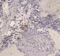
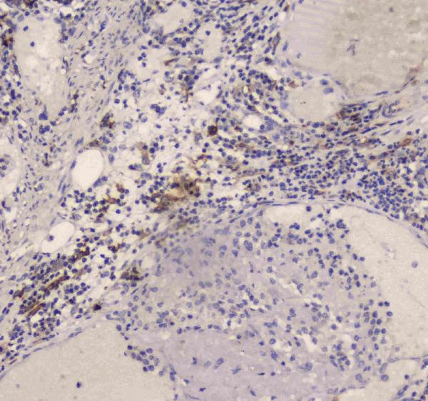
ARG43211 anti-CD209 / DC-SIGN antibody IHC-P image
Immunohistochemistry: Paraffin-embedded Human intestinal cancer tissue. Antigen Retrieval: Heat mediation was performed in Citrate buffer (pH 6.0) for 20 min. The tissue section was blocked with 10% goat serum. The tissue section was then stained with ARG43211 anti-CD209 / DC-SIGN antibody at 1 µg/ml dilution, overnight at 4°C.

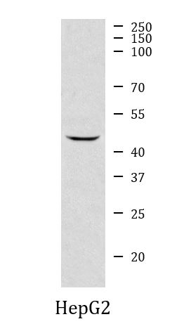
ARG43211 anti-CD209 / DC-SIGN antibody WB image
Western blot: 30 µg of sample under reducing conditions. HepG2 whole cell lysate stained with ARG43211 anti-CD209 / DC-SIGN antibody at 0.5 µg/ml dilution, overnight at 4°C.
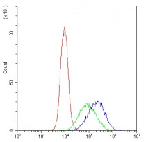
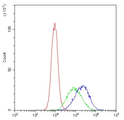
ARG43211 anti-CD209 / DC-SIGN antibody FACS image
Flow Cytometry: THP-1 cells were blocked with 10% normal goat serum and then stained with ARG43211 anti-CD209 / DC-SIGN antibody (blue) at 1 µg/10^6 cells for 30 min at 20°C, followed by incubation with DyLight®488 labelled secondary antibody. Isotype control antibody (green) was rabbit IgG (1 µg/10^6 cells) used under the same conditions. Unlabelled sample (red) was also used as a control.
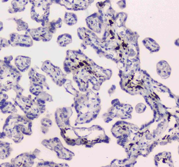
ARG43211 anti-CD209 / DC-SIGN antibody IHC-P image
Immunohistochemistry: Paraffin-embedded Human placenta tissue. Antigen Retrieval: Heat mediation was performed in Citrate buffer (pH 6.0) for 20 min. The tissue section was blocked with 10% goat serum. The tissue section was then stained with ARG43211 anti-CD209 / DC-SIGN antibody at 1 µg/ml dilution, overnight at 4°C.


 New Products


