


anti-CD274 / PD-L1 antibody


Key features and details
- 产品描述:
- 反应物种:
- 应用:
- 宿主:
- 克隆:
- 同位型:
- 靶点名称:
- 抗原物种:
- 抗原:
-
Brand:

 Product Details
| 产品描述 | Goat Polyclonal antibody recognizes CD274 / PD-L1 |
|---|---|
| 反应物种 | Hu |
| 应用 | FACS, ICC/IF, IHC-P, WB |
| 宿主 | Goat |
| 克隆 | Polyclonal |
| 同位型 | IgG |
| 靶点名称 | CD274 / PD-L1 |
| 抗原物种 | Human |
| 抗原 | CKKQSDTHLEET |
| 偶联标记 | Un-conjugated |
| 別名 | Programmed cell death 1 ligand 1; B7-H1; B7H1; PDL1; PDCD1 ligand 1; B7 homolog 1; PD-L1; CD antigen CD274; PDCD1L1; B7-H; Programmed death ligand 1; PDCD1LG1 |
| 应用建议 |
| ||||||||||
|---|---|---|---|---|---|---|---|---|---|---|---|
| 应用说明 | WB: Recommend incubate at RT for 1h. IHC-P: Antigen Retrieval: (1) Microwaved tissue section in Citrate buffer (pH 6.0), or (2) Steam tissue section in Tris/EDTA buffer (pH 9.0). * The dilutions indicate recommended starting dilutions and the optimal dilutions or concentrations should be determined by the scientist. | ||||||||||
| 阳性对照 | Human heart and U2OS | ||||||||||
| 实际分子量 | ~ 50 kDa |
| 形式 | Liquid |
|---|---|
| 纯化 | Purified from goat serum by ammonium sulphate precipitation followed by antigen affinity chromatography using the immunizing peptide. |
| 缓冲液 | Tris saline (pH 7.3), 0.02% Sodium azide and 0.5% BSA |
| 抗菌剂 | 0.02% Sodium azide |
| 稳定剂 | 0.5% BSA |
| 浓度 | 0.5 mg/ml |
| 存放说明 | For continuous use, store undiluted antibody at 2-8°C for up to a week. For long-term storage, aliquot and store at -20°C or below. Storage in frost free freezers is not recommended. Avoid repeated freeze/thaw cycles. Suggest spin the vial prior to opening. The antibody solution should be gently mixed before use. |
| 注意事项 | For laboratory research only, not for drug, diagnostic or other use. |
| 数据库连接 | |
|---|---|
| 基因名称 | CD274 |
| 全名 | CD274 molecule |
| 背景介绍 | CD274 / PD-L1 is an immune inhibitory receptor ligand. It is expressed by hematopoietic and non-hematopoietic cells, such as T cells and B cells and various types of tumor cells. The encoded protein is a type I transmembrane protein that has immunoglobulin V-like and C-like domains. Interaction of this ligand with its receptor inhibits T-cell activation and cytokine production. During infection or inflammation of normal tissue, this interaction is important for preventing autoimmunity by maintaining homeostasis of the immune response. In tumor microenvironments, this interaction provides an immune escape for tumor cells through cytotoxic T-cell inactivation. Expression of this gene in tumor cells is considered to be prognostic in many types of human malignancies, including colon cancer and renal cell carcinoma. Alternative splicing results in multiple transcript variants. [provided by RefSeq, Sep 2015] |
| 生物功能 | CD274 / PD-L1 plays a critical role in induction and maintenance of immune tolerance to self (PubMed:11015443, PubMed:28813417, PubMed:28813410). As a ligand for the inhibitory receptor PDCD1/PD-1, modulates the activation threshold of T-cells and limits T-cell effector response (PubMed:11015443, PubMed:28813417, PubMed:28813410). Through a yet unknown activating receptor, may costimulate T-cell subsets that predominantly produce interleukin-10 (IL10) (PubMed:10581077). The PDCD1-mediated inhibitory pathway is exploited by tumors to attenuate anti-tumor immunity and escape destruction by the immune system, thereby facilitating tumor survival (PubMed:28813417, PubMed:28813410). The interaction with PDCD1/PD-1 inhibits cytotoxic T lymphocytes (CTLs) effector function. The blockage of the PDCD1-mediated pathway results in the reversal of the exhausted T-cell phenotype and the normalization of the anti-tumor response, providing a rationale for cancer immunotherapy. [UniProt] |
| 细胞定位 | Cell membrane and Endomembrane system. |
| 产品亮点 | Related products: PD-L1 antibodies; PD-L1 ELISA Kits; Anti-Goat IgG secondary antibodies; Related news: The best solution for PD-1/PD-L1 research Examining CTL/NK-mediated cytotoxicity by ELISA |
| 研究领域 | Immune System antibody |
| 预测分子量 | 33 kDa |
ARG63715 anti-CD274 / PD-L1 antibody ICC/IF image
Immunofluorescence: Paraformaldehyde fixed A431 cells permeabilized with 0.15% Triton. Cells were stained with ARG63715 anti-CD274 / PD-L1 antibody (green) at 10 µg/ml dilution for 1 hour. DAPI (blue) for nuclear staining. Negative control: Unimmunized goat IgG (green) at 10 µg/ml dilution.
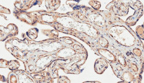
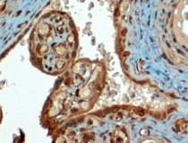
ARG63715 anti-CD274 / PD-L1 antibody IHC-P image
Immunohistochemistry: Paraffin-embedded Human placenta tissue. Antigen Retrieval: Microwaved tissue section in Citrate buffer (pH 6.0). The tissue section was stained with ARG63715 anti-CD274 / PD-L1 antibody at 2 µg/ml dilution followed by HRP-staining.
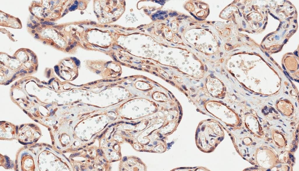
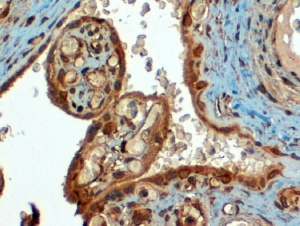
ARG63715 anti-CD274 / PD-L1 antibody IHC-P image
Immunohistochemistry: Paraffin-embedded Human placenta tissue. Antigen Retrieval: Steam tissue section in Tris/EDTA buffer (pH 9.0). The tissue section was stained with ARG63715 anti-CD274 / PD-L1 antibody at 4 µg/ml dilution followed by HRP-staining.

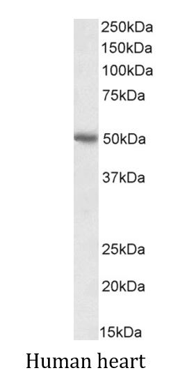
ARG63715 anti-CD274 / PD-L1 antibody WB image
Western blot: 35 µg of Human heart lysate (in RIPA buffer) stained with ARG63715 anti-CD274 / PD-L1 antibody at 0.01 µg/ml dilution and incubated at RT for 1 hour.
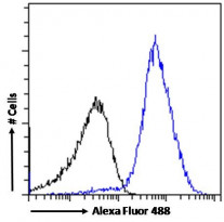
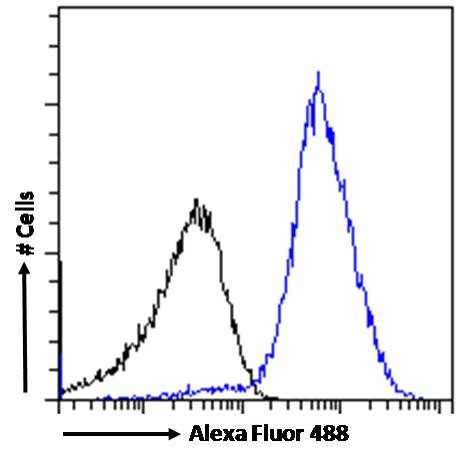
ARG63715 anti-CD274 / PD-L1 antibody FACS image
Flow Cytometry: Paraformaldehyde-fixed Jurkat cells permeabilized with 0.5% Triton. Cells were stained with ARG63715 anti-CD274 / PD-L1 antibody (blue line) at 10 µg/ml dilution for 1 hour, followed by incubation with Alexa Fluor 488 labelled secondary antibody. IgG control: Unimmunized goat IgG (black line).
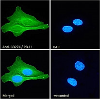
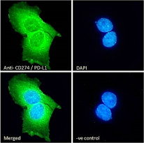
ARG63715 anti-CD274 / PD-L1 antibody ICC/IF image
Immunofluorescence: Paraformaldehyde fixed U2OS cells permeabilized with 0.15% Triton. Cells were stained with ARG63715 anti-CD274 / PD-L1 antibody (green) at 10 µg/ml dilution for 1 hour. DAPI (blue) for nuclear staining. Negative control: Unimmunized goat IgG (green) at 10 µg/ml dilution.
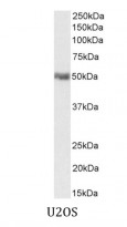
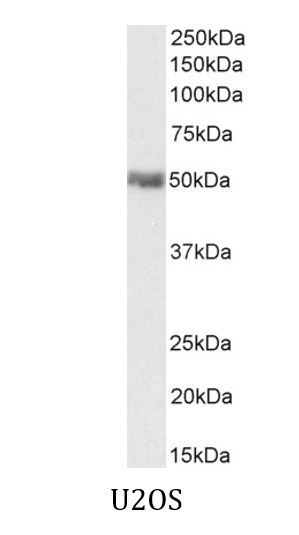
ARG63715 anti-CD274 / PD-L1 antibody WB image
Western blot: 35 µg of U2OS cell lysate (in RIPA buffer) stained with ARG63715 anti-CD274 / PD-L1 antibody at 0.1 µg/ml dilution and incubated at RT for 1 hour.


 New Products


