


anti-CD274 / PD-L1 antibody [SQab1716]
![anti-CD274 / PD-L1 antibody [SQab1716]](/upload/image/products/ARG65862_IF_293.jpg)
![anti-CD274 / PD-L1 antibody [SQab1716]](/upload/image/products/ARG65862_FACS_293.jpg)
![anti-CD274 / PD-L1 antibody [SQab1716]](/upload/image/products/ARG65862_IHC_Hu_placenta.jpg)
![anti-CD274 / PD-L1 antibody [SQab1716]](/upload/image/products/ARG65862_IP_293.jpg)
![anti-CD274 / PD-L1 antibody [SQab1716]](/upload/image/products/ARG65862_IHC_Hu_hepatocellular-carcinoma.jpg)
![anti-CD274 / PD-L1 antibody [SQab1716]](/upload/image/products/ARG65862_IHC_Hu_lung-adenocarcinoma.jpg)
Key features and details
- 产品描述:
- 反应物种:
- 应用:
- 宿主:
- 克隆:
- 克隆号:
- 同位型:
- 靶点名称:
- 抗原物种:
-
Brand:

 Product Details
| 产品描述 | Recombinant Rabbit Monoclonal antibody [SQab1716] recognizes CD274 / PD-L1 |
|---|---|
| 反应物种 | Hu |
| 应用 | FACS, ICC/IF, IHC-P, IP |
| 宿主 | Rabbit |
| 克隆 | Monoclonal |
| 克隆号 | SQab1716 |
| 同位型 | IgG |
| 靶点名称 | CD274 / PD-L1 |
| 抗原物种 | Human |
| 抗原 | Recombinant protein around aa. 19-239 (extracellular) of Human CD274 / PD-L1. |
| 偶联标记 | Un-conjugated |
| 別名 | Programmed cell death 1 ligand 1; B7-H1; B7H1; PDL1; PDCD1 ligand 1; B7 homolog 1; PD-L1; CD antigen CD274; PDCD1L1; B7-H; Programmed death ligand 1; PDCD1LG1 |
| 应用建议 |
| ||||||||||
|---|---|---|---|---|---|---|---|---|---|---|---|
| 应用说明 | IHC-P: Antigen Retrieval: Boil tissue section in Tris/EDTA buffer (pH 9.0). * The dilutions indicate recommended starting dilutions and the optimal dilutions or concentrations should be determined by the scientist. |
| 形式 | Liquid |
|---|---|
| 纯化 | Purification with Protein A. |
| 缓冲液 | PBS, 0.01% Sodium azide, 40% Glycerol and 0.05% BSA. |
| 抗菌剂 | 0.01% Sodium azide |
| 稳定剂 | 40% Glycerol and 0.05% BSA |
| 存放说明 | For continuous use, store undiluted antibody at 2-8°C for up to a week. For long-term storage, aliquot and store at -20°C. Storage in frost free freezers is not recommended. Avoid repeated freeze/thaw cycles. Suggest spin the vial prior to opening. The antibody solution should be gently mixed before use. |
| 注意事项 | For laboratory research only, not for drug, diagnostic or other use. |
| 数据库连接 | |
|---|---|
| 基因名称 | CD274 |
| 全名 | CD274 molecule |
| 背景介绍 | CD274 / PD-L1 is an immune inhibitory receptor ligand. It is expressed by hematopoietic and non-hematopoietic cells, such as T cells and B cells and various types of tumor cells. The encoded protein is a type I transmembrane protein that has immunoglobulin V-like and C-like domains. Interaction of this ligand with its receptor inhibits T-cell activation and cytokine production. During infection or inflammation of normal tissue, this interaction is important for preventing autoimmunity by maintaining homeostasis of the immune response. In tumor microenvironments, this interaction provides an immune escape for tumor cells through cytotoxic T-cell inactivation. Expression of this gene in tumor cells is considered to be prognostic in many types of human malignancies, including colon cancer and renal cell carcinoma. Alternative splicing results in multiple transcript variants. [provided by RefSeq, Sep 2015] |
| 生物功能 | CD274 / PD-L1 plays a critical role in induction and maintenance of immune tolerance to self (PubMed:11015443, PubMed:28813417, PubMed:28813410). As a ligand for the inhibitory receptor PDCD1/PD-1, modulates the activation threshold of T-cells and limits T-cell effector response (PubMed:11015443, PubMed:28813417, PubMed:28813410). Through a yet unknown activating receptor, may costimulate T-cell subsets that predominantly produce interleukin-10 (IL10) (PubMed:10581077). The PDCD1-mediated inhibitory pathway is exploited by tumors to attenuate anti-tumor immunity and escape destruction by the immune system, thereby facilitating tumor survival (PubMed:28813417, PubMed:28813410). The interaction with PDCD1/PD-1 inhibits cytotoxic T lymphocytes (CTLs) effector function. The blockage of the PDCD1-mediated pathway results in the reversal of the exhausted T-cell phenotype and the normalization of the anti-tumor response, providing a rationale for cancer immunotherapy. [UniProt] |
| 细胞定位 | Cell membrane and Endomembrane system. |
| 产品亮点 | Related products: PD-L1 antibodies; PD-L1 ELISA Kits; Anti-Rabbit IgG secondary antibodies; Related news: New PD-1 ELISA Kit, excellent for preclinical studies or pharmatheutical development Time to fight cancer by NK cells The best solution for PD-1/PD-L1 research Examining CTL/NK-mediated cytotoxicity by ELISA |
| 预测分子量 | 33 kDa |
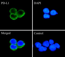
ARG65862 anti-CD274 / PD-L1 antibody [SQab1716] ICC/IF image
Immunofluorescence: 293 cells trasnfected with PD-L1 gene. Cells were fixed with 4% paraformaldehyde for 30 min at RT, permeabilized with 0.1% Triton X-100 for 10 min at RT then blocked with 10% goat serum for half an hour at room temperature. Samples were stained with ARG65862 anti-CD274 / PD-L1 antibody [SQab1716] (green) at 1:10000 at 4°C. DAPI (blue) was used as the nuclear counter stain.
Control: PBS and secondary antibody.
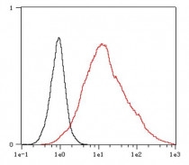
ARG65862 anti-CD274 / PD-L1 antibody [SQab1716] FACS image
Flow Cytometry: 293 cells transfected with PD-L1 gene and stained with ARG65862 anti-CD274 / PD-L1 antibody [SQab1716] at 1:200, in 1x PBS/1% BSA for 30 min at room temperature, followed by Alexa Fluor® 488 labelled secondary antibody (red histogram). Unlabelled sample (black histogram) was used as a control.
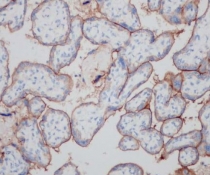
ARG65862 anti-CD274 / PD-L1 antibody [SQab1716] IHC-P image
Immunohistochemistry: Formalin/PFA-fixed and paraffin-embedded sections of Human placenta tissue stained with ARG65862 anti-CD274 / PD-L1 antibody [SQab1716] at 1:200 dilution. Antigen Retrieval: Boil tissue section in Tris/EDTA buffer (pH 9.0).
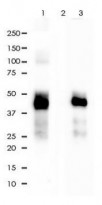
ARG65862 anti-CD274 / PD-L1 antibody [SQab1716] IP image
Immunoprecipitation: PD-L1 was immunoprecipitated from 0.2 mg of PD-L1 gene transfected 293 whole cells lysate by ARG65862 anti-CD274 / PD-L1 antibody [SQab1716] at 1:30 dilution. The immunoprecipitated lysates were then stained with ARG65862 anti-CD274 / PD-L1 antibody [SQab1716] by WB.
Lane 1: ARG65862 IP in 293 whole cell lysate transfected with PD-L1 gene.
Lane 2: PBS instead of ARG65862 in 293 whole cell lysate transfected with PD-L1 gene.
Lane 3: 293 whole cell lysate transfected with PD-L1 gene, 2 μg (input).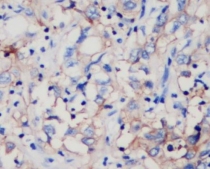
ARG65862 anti-CD274 / PD-L1 antibody [SQab1716] IHC-P image
Immunohistochemistry: Formalin/PFA-fixed and paraffin-embedded sections of Human hepatocellular carcinoma tissue stained with ARG65862 anti-CD274 / PD-L1 antibody [SQab1716] at 1:200 dilution. Antigen Retrieval: Boil tissue section in Tris/EDTA buffer (pH 9.0).
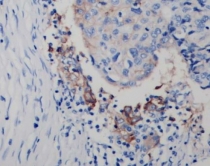
ARG65862 anti-CD274 / PD-L1 antibody [SQab1716] IHC-P image
Immunohistochemistry: Formalin/PFA-fixed and paraffin-embedded sections of Human lung adenocarcinoma tissue stained with ARG65862 anti-CD274 / PD-L1 antibody [SQab1716] at 1:200 dilution. Antigen Retrieval: Boil tissue section in Tris/EDTA buffer (pH 9.0).


Glucose Starvation Promotes anti-PD-L1 and anti-GLUT1 Activity of Metformin on Skin Cancer Cells
FACS / Human


Comparative transcriptomic analysis reveals differences in gene expression and regulatory pathways between nonacral and acral melanoma in Asian individuals
IHC-P / Human
Human Metastatic Melanoma Cell Lines Panel for In Vitro and In Vivo Investigations
FACS / Human
Quantitative Immunofluorescence Evaluation of PD-L1 Expression in Non-Muscle-Invasive and Muscle-Invasive Urothelial Bladder Cancer
IHC-P / Human
ПАНЕЛЬ КУЛЬТУР ОПУХОЛЕВЫХ КЛЕТОК С ОХАРАКТЕРИЗОВАННОЙ ЭКСПРЕССИЕЙ БЕЛКА PD-L1 ДЛЯ ДОКЛИНИЧЕСКОЙ ОЦЕНКИ ВЗАИМОДЕЙСТВИЯ ПРОТИВООПУХОЛЕВЫХ ПРЕПАРАТОВ С ИНГИБИТОРАМИ КОНТРОЛЬНЫХ ТОЧЕК ИММУНИТЕТА
FACS / Human
Количественная иммунофлуоресцентная оценка показателей экспрессии PD-L1 в ткани немышечно-инвазивного и мышечно-инвазивного уротелиального рака мочевого пузыря
ICC/IF / Human
New Products


