


anti-CD2AP antibody


Key features and details
- 产品描述:
- 反应物种:
- 应用:
- 宿主:
- 克隆:
- 同位型:
- 靶点名称:
- 抗原物种:
- 抗原:
-
Brand:

 Product Details
| 产品描述 | Rabbit Polyclonal antibody recognizes CD2AP |
|---|---|
| 反应物种 | Hu, Ms, Rat |
| 应用 | FACS, ICC/IF, IHC-P, WB |
| 宿主 | Rabbit |
| 克隆 | Polyclonal |
| 同位型 | IgG |
| 靶点名称 | CD2AP |
| 抗原物种 | Human |
| 抗原 | Recombinant protein corresponding to K253-K337 of Human CD2AP. |
| 偶联标记 | Un-conjugated |
| 別名 | Adapter protein CMS; CD2-associated protein; CMS; Cas ligand with multiple SH3 domains |
| 应用建议 |
| ||||||||||
|---|---|---|---|---|---|---|---|---|---|---|---|
| 应用说明 | IHC-P: Antigen Retrieval: Heat mediation was performed in Citrate buffer (pH 6.0) for 20 min. * The dilutions indicate recommended starting dilutions and the optimal dilutions or concentrations should be determined by the scientist. |
| 形式 | Liquid |
|---|---|
| 纯化 | Affinity purification with immunogen. |
| 缓冲液 | 0.9% NaCl, 0.2% Na2HPO4, 0.05% Sodium azide and 4% Trehalose. |
| 抗菌剂 | 0.05% Sodium azide |
| 稳定剂 | 4% Trehalose |
| 浓度 | 0.5 mg/ml |
| 存放说明 | For continuous use, store undiluted antibody at 2-8°C for up to a week. For long-term storage, aliquot and store at -20°C or below. Storage in frost free freezers is not recommended. Avoid repeated freeze/thaw cycles. Suggest spin the vial prior to opening. The antibody solution should be gently mixed before use. |
| 注意事项 | For laboratory research only, not for drug, diagnostic or other use. |
| 数据库连接 | |
|---|---|
| 基因名称 | CD2AP |
| 全名 | CD2-associated protein |
| 背景介绍 | This gene encodes a scaffolding molecule that regulates the actin cytoskeleton. The protein directly interacts with filamentous actin and a variety of cell membrane proteins through multiple actin binding sites, SH3 domains, and a proline-rich region containing binding sites for SH3 domains. The cytoplasmic protein localizes to membrane ruffles, lipid rafts, and the leading edges of cells. It is implicated in dynamic actin remodeling and membrane trafficking that occurs during receptor endocytosis and cytokinesis. Haploinsufficiency of this gene is implicated in susceptibility to glomerular disease. [provided by RefSeq, Jul 2008] |
| 生物功能 | Seems to act as an adapter protein between membrane proteins and the actin cytoskeleton. In collaboration with CBLC, modulates the rate of RET turnover and may act as regulatory checkpoint that limits the potency of GDNF on neuronal survival. Controls CBLC function, converting it from an inhibitor to a promoter of RET degradation. May play a role in receptor clustering and cytoskeletal polarity in the junction between T-cell and antigen-presenting cell. May anchor the podocyte slit diaphragm to the actin cytoskeleton in renal glomerolus. Also required for cytokinesis. [UniProt] |
| 细胞定位 | Cytoplasm, cytoskeleton. Cell projection, ruffle. Cell junction. Note=Colocalizes with F-actin and BCAR1/p130Cas in membrane ruffles (PubMed:10339567). Located at podocyte slit diaphragm between podocyte foot processes (By similarity). During late anaphase and telophase, concentrates in the vicinity of the midzone microtubules and in the midbody in late telophase (PubMed:15800069). [UniProt] |
| 预测分子量 | 71 kDa |
| 翻译后修饰 | Phosphorylated on tyrosine residues; probably by c-Abl, Fyn and c-Src. [UniProt] |

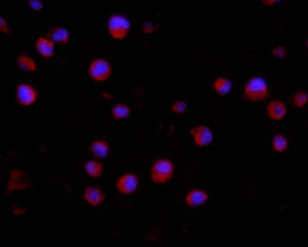
ARG59305 anti-CD2AP antibody ICC/IF image
Immunofluorescence: K562 cells were blocked with 10% goat serum and then stained with ARG59305 anti-CD2AP antibody (red) at 2 µg/ml dilution, overnight at 4°C. DAPI (blue) for nuclear staining.
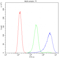
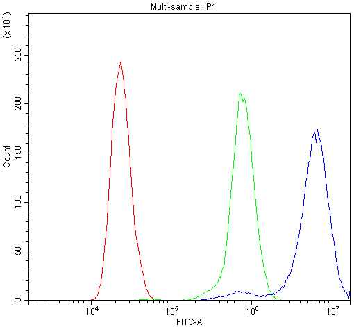
ARG59305 anti-CD2AP antibody FACS image
Flow Cytometry: A431 cells were blocked with 10% normal goat serum and then stained with ARG59305 anti-CD2AP antibody (blue) at 1 µg/10^6 cells for 30 min at 20°C, followed by DyLight®488 labelled secondary antibody. Isotype control antibody (green) was rabbit IgG (1 µg/10^6 cells) used under the same conditions. Unlabelled sample (red) was also used as a control.

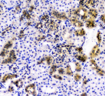
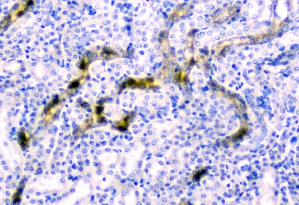
ARG59305 anti-CD2AP antibody IHC-P image
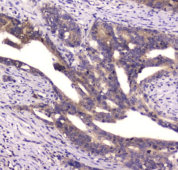
Immunohistochemistry: Paraffin-embedded Human intestinal cancer tissue. Antigen Retrieval: Heat mediation was performed in Citrate buffer (pH 6.0) for 20 min. The tissue section was blocked with 10% goat serum. The tissue section was then stained with ARG59305 anti-CD2AP antibody at 1 µg/ml dilution, overnight at 4°C.
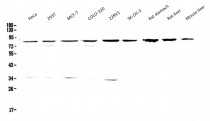
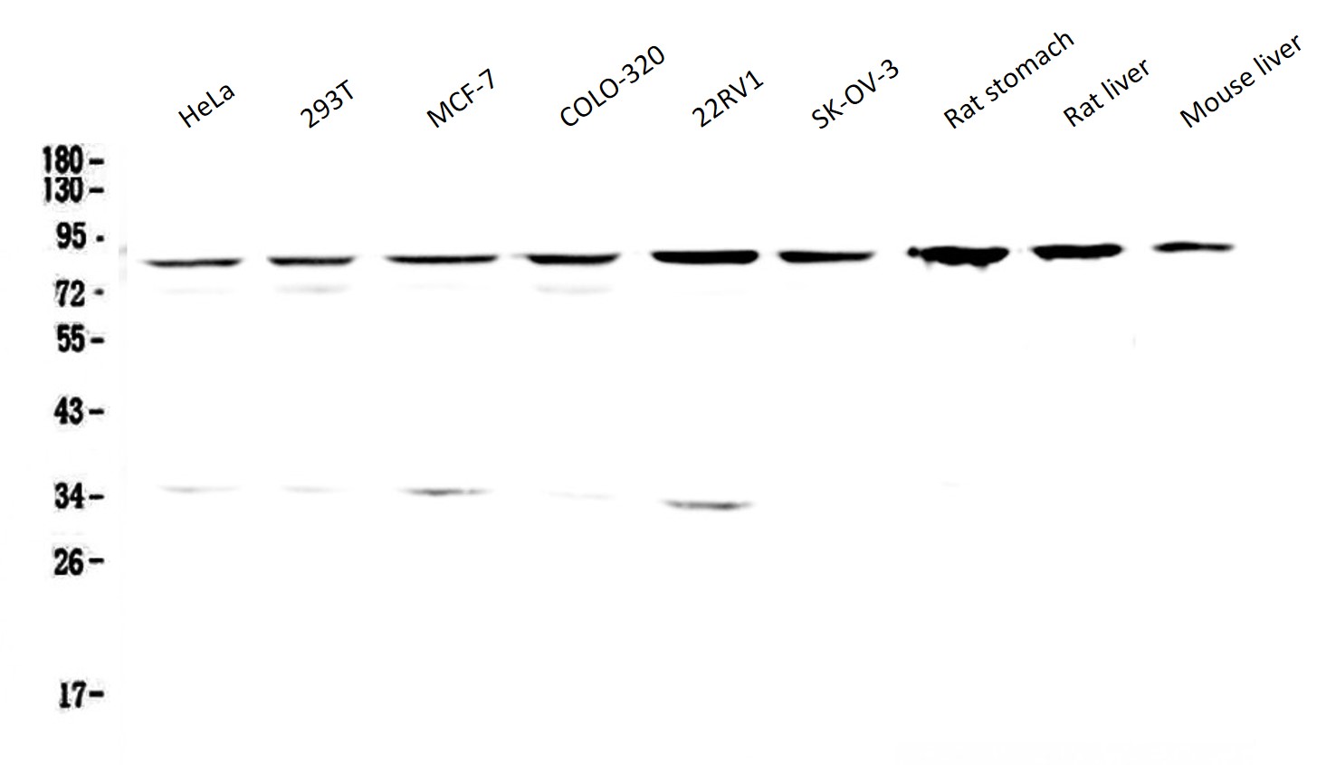
ARG59305 anti-CD2AP antibody WB image
Western blot: 50 µg of samples under reducing conditions. HeLa, 293T, MCF-7, COLO-320, 22RV1, SK-OV-3, Rat stomach, Rat liver and Mouse liver lysates stained with ARG59305 anti-CD2AP antibody at 0.5 µg/ml, overnight at 4°C.
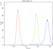
ARG59305 anti-CD2AP antibody FACS image
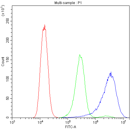
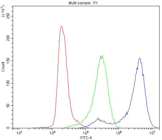
Flow Cytometry: Caco-2 cells were blocked with 10% normal goat serum and then stained with ARG59305 anti-CD2AP antibody (blue) at 1 µg/10^6 cells for 30 min at 20°C, followed by DyLight®488 labelled secondary antibody. Isotype control antibody (green) was rabbit IgG (1 µg/10^6 cells) used under the same conditions. Unlabelled sample (red) was also used as a control.
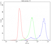
ARG59305 anti-CD2AP antibody FACS image
Flow Cytometry: K562 cells were blocked with 10% normal goat serum and then stained with ARG59305 anti-CD2AP antibody (blue) at 1 µg/10^6 cells for 30 min at 20°C, followed by DyLight®488 labelled secondary antibody. Isotype control antibody (green) was rabbit IgG (1 µg/10^6 cells) used under the same conditions. Unlabelled sample (red) was also used as a control.
ARG59305 anti-CD2AP antibody IHC-P image
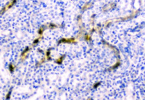
Immunohistochemistry: Paraffin-embedded Rat kidney tissue. Antigen Retrieval: Heat mediation was performed in Citrate buffer (pH 6.0) for 20 min. The tissue section was blocked with 10% goat serum. The tissue section was then stained with ARG59305 anti-CD2AP antibody at 1 µg/ml dilution, overnight at 4°C.
ARG59305 anti-CD2AP antibody IHC-P image
Immunohistochemistry: Paraffin-embedded Mouse kidney tissue. Antigen Retrieval: Heat mediation was performed in Citrate buffer (pH 6.0) for 20 min. The tissue section was blocked with 10% goat serum. The tissue section was then stained with ARG59305 anti-CD2AP antibody at 1 µg/ml dilution, overnight at 4°C.


 New Products


