


anti-CDC20 antibody


Key features and details
- 产品描述:
- 反应物种:
- 应用:
- 宿主:
- 克隆:
- 同位型:
- 靶点名称:
- 抗原物种:
- 抗原:
-
Brand:

 Product Details
| 产品描述 | Rabbit Polyclonal antibody recognizes CDC20 |
|---|---|
| 反应物种 | Hu, Ms, Rat |
| 应用 | FACS, ICC/IF, IHC-P, WB |
| 宿主 | Rabbit |
| 克隆 | Polyclonal |
| 同位型 | IgG |
| 靶点名称 | CDC20 |
| 抗原物种 | Human |
| 抗原 | Synthetic peptide corresponding to a sequence of Human CDC20. (QTPTKKEHQKAWALNLNGFDVEEAKILRLSGKPQNAPEGYQNRLKVLYSQKAT) |
| 偶联标记 | Un-conjugated |
| 別名 | CDC20A; p55CDC; Cell division cycle protein 20 homolog; bA276H19.3 |
| 应用建议 |
| ||||||||||
|---|---|---|---|---|---|---|---|---|---|---|---|
| 应用说明 | * The dilutions indicate recommended starting dilutions and the optimal dilutions or concentrations should be determined by the scientist. | ||||||||||
| 阳性对照 | HeLa | ||||||||||
| 实际分子量 | ~ 55 kDa |
| 形式 | Liquid |
|---|---|
| 纯化 | Affinity purification with immunogen. |
| 缓冲液 | 0.2% Na2HPO4, 0.9% NaCl, 0.05% Sodium azide and 4% Trehalose. |
| 抗菌剂 | 0.05% Sodium azide |
| 稳定剂 | 4% Trehalose |
| 浓度 | 0.5 mg/ml |
| 存放说明 | For continuous use, store undiluted antibody at 2-8°C for up to a week. For long-term storage, aliquot and store at -20°C or below. Storage in frost free freezers is not recommended. Avoid repeated freeze/thaw cycles. Suggest spin the vial prior to opening. The antibody solution should be gently mixed before use. |
| 注意事项 | For laboratory research only, not for drug, diagnostic or other use. |
| 数据库连接 | |
|---|---|
| 基因名称 | CDC20 |
| 全名 | cell division cycle 20 |
| 背景介绍 | CDC20 appears to act as a regulatory protein interacting with several other proteins at multiple points in the cell cycle. It is required for two microtubule-dependent processes, nuclear movement prior to anaphase and chromosome separation. [provided by RefSeq, Jul 2008] |
| 生物功能 | Required for full ubiquitin ligase activity of the anaphase promoting complex/cyclosome (APC/C) and may confer substrate specificity upon the complex. Is regulated by MAD2L1: in metaphase the MAD2L1-CDC20-APC/C ternary complex is inactive and in anaphase the CDC20-APC/C binary complex is active in degrading substrates. The CDC20-APC/C complex positively regulates the formation of synaptic vesicle clustering at active zone to the presynaptic membrane in postmitotic neurons. CDC20-APC/C-induced degradation of NEUROD2 induces presynaptic differentiation. [UniProt] |
| 细胞定位 | Cytoplasm, cytoskeleton, microtubule organizing center, centrosome. Cytoplasm, cytoskeleton, spindle pole. [UniProt] |
| 预测分子量 | 55 kDa |
| 翻译后修饰 | Acetylated. Deacetylated at Lys-66 by SIRT2; deacetylation enhances the interaction of CDC20 with CDC27, leading to activation of anaphase promoting complex/cyclosome (APC/C). Phosphorylated during mitosis, probably by maturation promoting factor (MPF). Phosphorylated by BUB1 at Ser-41; Ser-72; Ser-92; Ser-153; Thr-157 and Ser-161. Phosphorylated by NEK2. Dephosphorylated by CTDP1. Ubiquitinated and degraded by the proteasome during spindle assembly checkpoint. Deubiquitinated by USP44, leading to stabilize the MAD2L1-CDC20-APC/C ternary complex, thereby preventing premature activation of the APC/C. Ubiquitinated at Lys-490 during prometaphase. Ubiquitination at Lys-485 and Lys-490 has no effect on its ability to bind the APC/C complex. [UniProt] |

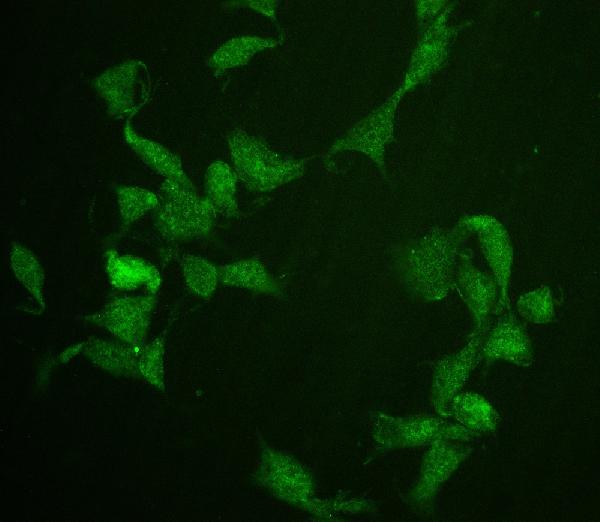
ARG42680 anti-CDC20 antibody ICC/IF image
Immunofluorescence: NIH/3T3 cells were blocked with 10% goat serum and then stained with ARG42680 anti-CDC20 antibody at 2 µg/ml dilution, overnight at 4°C.
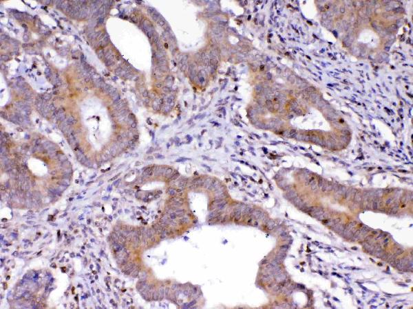
ARG42680 anti-CDC20 antibody IHC-P image
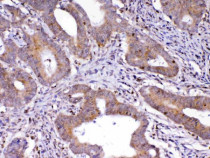
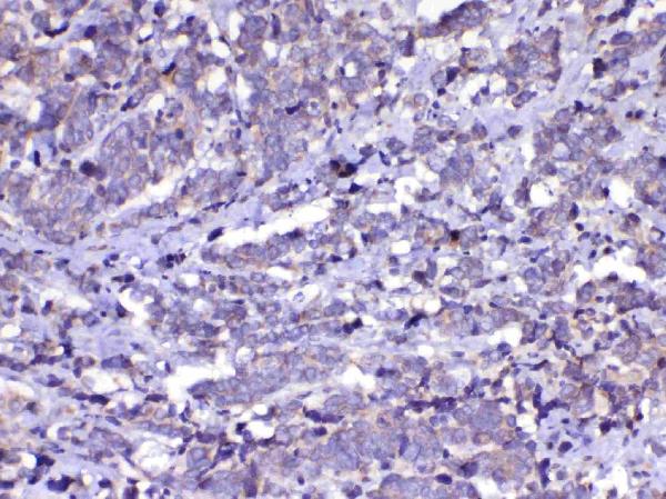
Immunohistochemistry: Paraffin-embedded Human colon cancer tissue. Antigen Retrieval: Heat mediation was performed in Citrate buffer (pH 6.0) for 20 min. The tissue section was blocked with 10% goat serum. The tissue section was then stained with ARG42680 anti-CDC20 antibody at 1 µg/ml dilution, overnight at 4°C.
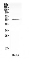
ARG42680 anti-CDC20 antibody WB image
Western blot: 50 µg of sample under reducing condition. HeLa whole cell lysate stained with ARG42680 anti-CDC20 antibody at 0.5 µg/ml dilution, overnight at 4°C.
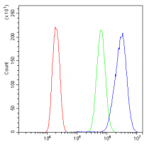
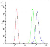
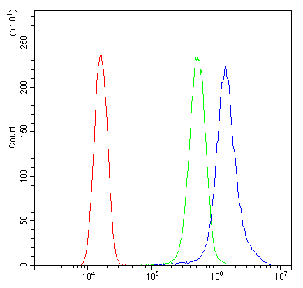
ARG42680 anti-CDC20 antibody FACS image
Flow Cytometry: U2OS cells were blocked with 10% normal goat serum and then stained with ARG42680 anti-CDC20 antibody (blue) at 1 µg/10^6 cells for 30 min at 20°C, followed by incubation with DyLight®488 labelled secondary antibody. Isotype control antibody (green) was rabbit IgG (1 µg/10^6 cells) used under the same conditions. Unlabelled sample (red) was also used as a control.
ARG42680 anti-CDC20 antibody IHC-P image
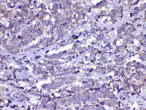
Immunohistochemistry: Paraffin-embedded Human lung cancer tissue. Antigen Retrieval: Heat mediation was performed in Citrate buffer (pH 6.0) for 20 min. The tissue section was blocked with 10% goat serum. The tissue section was then stained with ARG42680 anti-CDC20 antibody at 1 µg/ml dilution, overnight at 4°C.
ARG42680 anti-CDC20 antibody IHC-P image
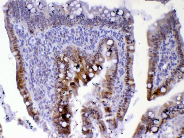
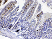
Immunohistochemistry: Paraffin-embedded Mouse small intestine tissue. Antigen Retrieval: Heat mediation was performed in Citrate buffer (pH 6.0) for 20 min. The tissue section was blocked with 10% goat serum. The tissue section was then stained with ARG42680 anti-CDC20 antibody at 1 µg/ml dilution, overnight at 4°C.

ARG42680 anti-CDC20 antibody IHC-P image
Immunohistochemistry: Paraffin-embedded Rat small intestine tissue. Antigen Retrieval: Heat mediation was performed in Citrate buffer (pH 6.0) for 20 min. The tissue section was blocked with 10% goat serum. The tissue section was then stained with ARG42680 anti-CDC20 antibody at 1 µg/ml dilution, overnight at 4°C.
ARG42680 anti-CDC20 antibody IHC-P image
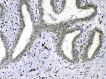
Immunohistochemistry: Paraffin-embedded Human mammary cancer tissue. Antigen Retrieval: Heat mediation was performed in Citrate buffer (pH 6.0) for 20 min. The tissue section was blocked with 10% goat serum. The tissue section was then stained with ARG42680 anti-CDC20 antibody at 1 µg/ml dilution, overnight at 4°C.
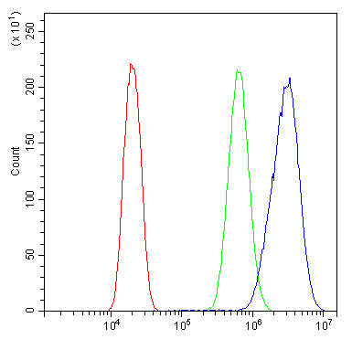
ARG42680 anti-CDC20 antibody FACS image
Flow Cytometry: SiHa cells were blocked with 10% normal goat serum and then stained with ARG42680 anti-CDC20 antibody (blue) at 1 µg/10^6 cells for 30 min at 20°C, followed by incubation with DyLight®488 labelled secondary antibody. Isotype control antibody (green) was rabbit IgG (1 µg/10^6 cells) used under the same conditions. Unlabelled sample (red) was also used as a control.


 New Products


