


anti-Cystatin B / Stefin B antibody


Key features and details
- 产品描述:
- 反应物种:
- 应用:
- 宿主:
- 克隆:
- 同位型:
- 靶点名称:
- 抗原物种:
- 抗原:
-
Brand:

 Product Details
| 产品描述 | Rabbit Polyclonal antibody recognizes Cystatin B / Stefin B |
|---|---|
| 反应物种 | Hu |
| 应用 | FACS, ICC/IF, IHC-P, WB |
| 宿主 | Rabbit |
| 克隆 | Polyclonal |
| 同位型 | IgG |
| 靶点名称 | Cystatin B / Stefin B |
| 抗原物种 | Human |
| 抗原 | Recombinant protein corresponding to M1-F98 Human Stefin B. |
| 偶联标记 | Un-conjugated |
| 別名 | Liver thiol proteinase inhibitor; EPM1; CPI-B; EPM1A; Cystatin-B; Stefin-B; PME; CST6; ULD; STFB |
| 应用建议 |
| ||||||||||
|---|---|---|---|---|---|---|---|---|---|---|---|
| 应用说明 | IHC-P: Antigen Retrieval: By heat mediation. * The dilutions indicate recommended starting dilutions and the optimal dilutions or concentrations should be determined by the scientist. |
| 形式 | Liquid |
|---|---|
| 纯化 | Affinity purification with immunogen. |
| 缓冲液 | 0.9% NaCl, 0.2% Na2HPO4, 0.05% Sodium azide and 5% BSA. |
| 抗菌剂 | 0.05% Sodium azide |
| 稳定剂 | 5% BSA |
| 浓度 | 0.5 mg/ml |
| 存放说明 | For continuous use, store undiluted antibody at 2-8°C for up to a week. For long-term storage, aliquot and store at -20°C or below. Storage in frost free freezers is not recommended. Avoid repeated freeze/thaw cycles. Suggest spin the vial prior to opening. The antibody solution should be gently mixed before use. |
| 注意事项 | For laboratory research only, not for drug, diagnostic or other use. |
| 数据库连接 | |
|---|---|
| 基因名称 | CSTB |
| 全名 | cystatin B (stefin B) |
| 背景介绍 | The cystatin superfamily encompasses proteins that contain multiple cystatin-like sequences. Some of the members are active cysteine protease inhibitors, while others have lost or perhaps never acquired this inhibitory activity. There are three inhibitory families in the superfamily, including the type 1 cystatins (stefins), type 2 cystatins and kininogens. This gene encodes a stefin that functions as an intracellular thiol protease inhibitor. The protein is able to form a dimer stabilized by noncovalent forces, inhibiting papain and cathepsins l, h and b. The protein is thought to play a role in protecting against the proteases leaking from lysosomes. Evidence indicates that mutations in this gene are responsible for the primary defects in patients with progressive myoclonic epilepsy (EPM1). [provided by RefSeq, Jul 2008] |
| 生物功能 | This is an intracellular thiol proteinase inhibitor. Tightly binding reversible inhibitor of cathepsins L, H and B. [UniProt] |
| 细胞定位 | Cytoplasm. Nucleus. [UniProt] |
| 预测分子量 | 11 kDa |
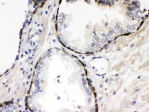
ARG59680 anti-Cystatin B / Stefin B antibody IHC-P image
Immunohistochemistry: Paraffin-embedded Human prostatic cancer stained with ARG59680 anti-Cystatin B / Stefin B antibody.
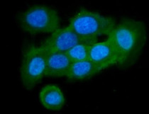
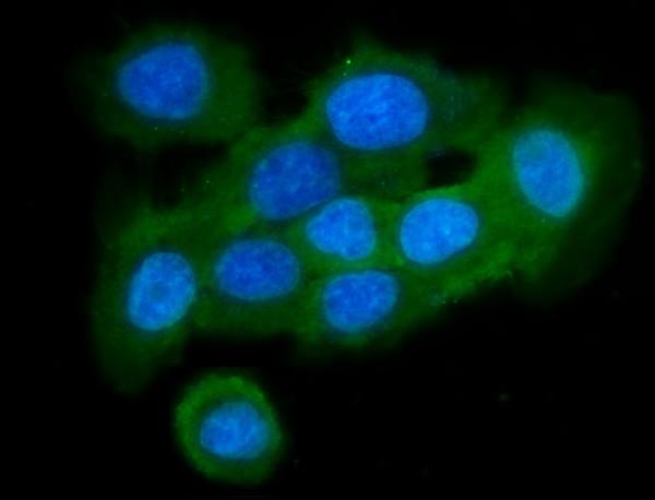
ARG59680 anti-Cystatin B / Stefin B antibody ICC/IF image
Immunofluorescence: A431 cells stained with ARG59680 anti-Cystatin B / Stefin B antibody at 1:100 dilution.
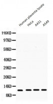
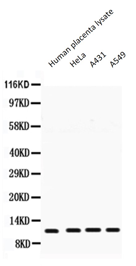
ARG59680 anti-Cystatin B / Stefin B antibody WB image
Western blot: 50 µg of Human placenta lysate, 40 µg of HeLa whole cell lysate, 40 µg of A431 whole cell lysate and 40 µg of A549 whole cell lysate stained with ARG59680 anti-Cystatin B / Stefin B antibody at 0.5 µg/ml dilution.
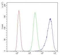
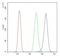
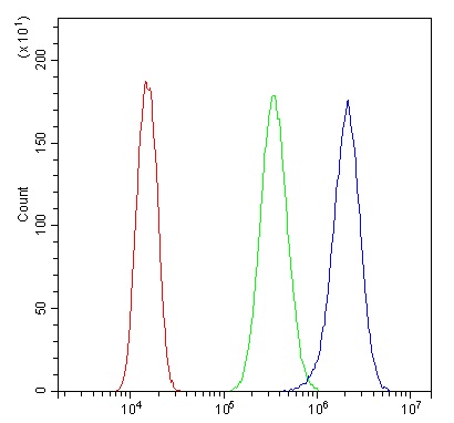
ARG59680 anti-Cystatin B / Stefin B antibody FACS image
Flow Cytometry: A431 cells were blocked with 10% normal goat serum and then stained with ARG59680 anti-Cystatin B / Stefin B antibody (blue) at 1 µg/10^6 cells for 30 min at 20°C, followed by incubation with DyLight®488 labelled secondary antibody. Isotype control antibody (green) was rabbit IgG (1 µg/10^6 cells) used under the same conditions. Unlabelled sample (red) was also used as a control.
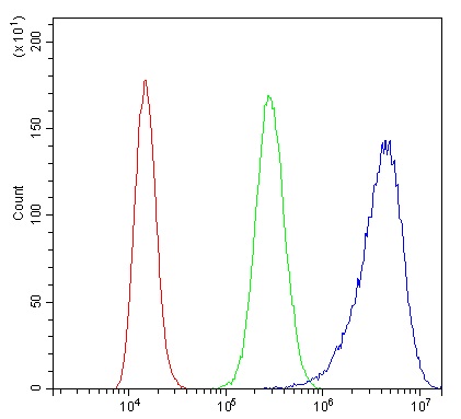
ARG59680 anti-Cystatin B / Stefin B antibody FACS image
Flow Cytometry: U2OS cells were blocked with 10% normal goat serum and then stained with ARG59680 anti-Cystatin B / Stefin B antibody (blue) at 1 µg/10^6 cells for 30 min at 20°C, followed by incubation with DyLight®488 labelled secondary antibody. Isotype control antibody (green) was rabbit IgG (1 µg/10^6 cells) used under the same conditions. Unlabelled sample (red) was also used as a control.


 New Products


